INTRODUCTION
Cervical spondylotic myelopathy (CSM) is one of the most common disorders of the cervical spine requiring surgical treatment. It is characterized by development and progression of degenerative changes associated with normal aging process. Cervical spinal stenosis patients have a tendency to suffer chronic myelopathy and high risk of acute spinal cord injury after traumatic injury. Mostly, cervical canal compromise result from two types of spinal degenerative processes, (1) ventral cord compression from bulging discs and osteophytes and (2) posterior compression by facet hypertrophy and thickening of the ligamenta flavum. The average anteroposterior diameter of a normal cervical canal on plain radiograph is 17mm, whereas symptomatic stenosis generally occurs when the diameter is less than 13mm33).
Various anterior and posterior surgical approaches for the treatment of CSM have been introduced, and studies have shown differing results depending on approaches8,16,20-22,24,25,28). Concerning the disadvantages of detaching the cervical paraspinal muscles from the laminae and the spinous processes in conventional posterior approaches7,26), anterior approach has become dominant among spinal surgeons, worldwide. Surgical trauma to the extensor cervical muscles in conventional posterior cervical operation is a major cause of various postoperative complications2,14,18,36), such as persistent neck and shoulder pain, postoperative kyphosis, spinal instability, etc. The current trend of favoring anterior approach surgery, even in patients with normal sagittal balance and posterior compressive pathology, is of great concern with the possibility of complications such as adjacent level disease and recurrent laryngeal and esophageal injury1,5,9,17).
Recently, with the increasing popularity of minimally invasive techniques, the possibility of posterior cervical approach is gaining renewed interest4,6,13). With the aid of a tubular retractor system, minimally invasive posterior cervical decompression has become possible, even in the multi-level cervical diseases. These evolutions of surgical techniques have led to a greater reduction in tissue damage during operation, which reduces postoperative pain, shortens hospital stays, quicker return to daily living activities.
The objective of this study is to introduce a novel minimally invasive surgical technique for multi-level posterior cervical decompression using a tubular retractor system and to evaluate safety and efficacy of this technique in short-term follow-up. This study is a series of consecutive mid-term follow-up reports in controlled clinical trials held at the institute of the authors using a minimally invasive surgical technique15).
MATERIALS AND METHODS
1. Materials and Methods
Twenty-one patients suffering from CSM underwent minimally invasive posterior cervical decompression using a tubular retractor. The operations were performed between April 2012 and May 2014. The indications for surgery were (1) presence of CSM confirmed by radiologic imaging studies, (2) presence of symptomatic myelopathy for more than 6 months, (3) compression ratio less than 0.4, indicating flattening of the spinal cord, (4) transverse area of the cord less than 40 mm2, (5) predominant dorsal cord compressing pathology such as ossification of the ligamenta flava (OLF), and (6) failure of conservative treatment over a period of 6 weeks. The exclusion criteria were cervical myelopathy with tumor, trauma, severe ossification of the posterior longitudinal ligament (OPLL), herniated disc, rheumatoid arthritis, pyogenic spondylitis, and the presence of other combined spinal lesions. The pathologic level and extent of spinal cord compression were confirmed by magnetic resonance imaging (MRI) and post-myelography computed tomography (CT). In addition, cervical MRI was performed in three different neck positions (neutral, flexion, and extension) in all patients to determine whether the spinal canal was dominantly compressed by posterior or anterior pathology. Patients with dominant anterior compression (such as multi-level intervertebral disc bulging) were excluded from the study and underwent surgery using an alternative anterior approach. The demographic and intraoperative data of the patients are listed in Table 1. The study included 9 men and 12 women. All patients presented with symptoms of cervical myelopathy: clumsiness, numbness of the upper and lower extremities, gait disturbances, urinary disturbances, etc. The average age of the study subjects at the time of operation was 56.7±14.1 years and the average body mass index was 26.3±3.4 kg/m2. The mean visual analogue scale (VAS) scores of preoperative neck pain and radicular arm pain were 6.4±2.7 and 8.9±2.1, respectively, and the average duration of pain was 17.4±8.7 months. One of the patients had undergone previous anterior cervical fusion at a local clinic for a herniated disc. Eight patients were operated on at one segment, four patients at two segments, and nine patients at three segments.
The hospital charts and follow-up medical records of all patients were carefully reviewed. Outcomes were assessed preoperatively and postoperatively using the Japanese Orthopedic Association scoring system for cervical myelopathy (C-JOA score)34), recovery rate as calculated by Hirabayashi’s method12,34), a modified version of the Oswestry Disability Index called the neck disability index (NDI)30,38), and VAS score for neck and radicular arm pain38).
All parameters were statistically analyzed. The data are expressed as mean±standard deviation (SD). A result was considered statistically significant if the p-value was less than 0.05.
2. Surgical Technique
Detailed surgical technique is introduced in our previous reports15).
Before surgery, each patient underwent evaluation by dynamic radiographs to rule out obvious instability, and MRI or post-myelography CT to define the necessary extent of the surgery. The results of routine medical and laboratory evaluations were obtained. The anesthesia team was informed prior to the surgery of the possible need for fiberoptic intubation. The operation was done under general anesthesia and monitoring of interoperative somatosensory evoked potentials (SEP) was done in all patients.
RESULTS
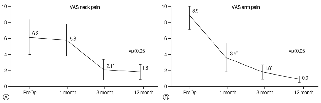
Twenty-one patients underwent minimally invasive posterior cervical decompression using a tubular retractor system and a surgical microscope. The mean follow-up duration was 13± 5.3 months. Average intraoperative blood loss was 61±33mL, average operating time was 73.3±21.4 min, and average length of hospital stay was 1.4±1.5 days (Table 1). Patients with sedentary jobs usually returned to work within a week after discharge. Muscle weakness improved in all patients. Sensory deficits resolved in 17 patients and improved in 4 patients. Analysis of the mean VAS scores for radicular pain and neck pain showed significant improvement compared to preoperative values at final follow-up (Fig. 1). The mean VAS score for neck pain decreased from 6.2±2.2 to 5.8±2.0 immediately post-operatively, reaching 2.1±1.3 at 3 months and 1.8±0.9 at the last follow-up visit. VAS scores for radicular arm pain also decreased, from 8.9±2.8 preoperatively to 3.6 ±1.4 immediately post-operatively, reaching 1.8±0.9 at 3 months and 0.9±0.4 at the last follow-up. Mean NDI decreased significantly, from 68.3±9.1 preoperatively to 13.3± 10.4 (p<0.05) at the final follow-up. Mean C-JOA score also improved from a preoperative value of 11.2±2.6 to 16.2±3.1 (p<0.05) at the last follow-up, and the recovery rate as calculated using the Hirabayashi method averaged 53.2±22.0% (Table 2). There were no significant operation-related complications such as cerebrospinal fluid (CSF) leakage, postoperative infection, instability, etc.
DISCUSSION
Selection of approach side for cervical spinal decompression usually depends on various factors, including extent of disease, sagittal curvature of the cervical spine, prior surgery, general condition of the patient, skill and familiarity of the surgeon, severity of canal compression, and intervertebral mobility at the level of maximum compression. The anterior approach offers a relatively simpler route to the spine and a means of decompressing the ventral spinal pathology than does the posterior approach. On the other hand, disadvantages of the anterior approach include potential complications involving anterior neck structures, dysphagia, recurrent laryngeal nerve injury, and adjacent segment degeneration following loss of one or more motion segments.
Posterior decompression allows safer route for the thecal sac and avoids many of the risks of anterior exposure. However, ventral compressing pathologies such as disc herniation, osteophytes, or OPLL may be neglected when using a posterior approach. Although a simple multilevel laminectomy or laminoplasty is a relatively straightforward procedure, it often results in significant postoperative neck pain and longer hospitalization. In addition, CSF leakage, wound-related problems, postoperative kyphosis, and instability are not uncommon in conventional posterior operations.
With the recent advancement of specialized surgical instruments and access devices, minimally invasive spinal surgery has proven to be a useful tool for the treatment of various spinal diseases while minimizing soft tissue damage. Application of this technique to the cervical spine followed naturally, and posterior minimally invasive cervical surgery has been performed recently in many institutions to determine the feasibility and efficacy of such procedures. Recent studies using a trans-muscular working channel to perform minimally invasive decompression for radiculopathy and myelopathy concluded that the basic technique is safe and feasible4,32).
There are a few reports of posterior cervical decompression using different minimally invasive techniques up to date. Routine three-position cervical MRI for cervical spondylotic myelopathic patients was performed in our series to evaluate the characteristics of canal compression and to aid in the surgeon’s selection of an appropriate surgical technique. Cervical dynamic MRI is useful in accurately determining the number of levels in which the spinal cord is compromised, and evaluating the degree of narrowing of the spinal canal3,37). Using radiologic information, we selected cases with more dorsal than ventral compression for this series. As mentioned above, individuals with more dominant anterior compression were excluded and underwent alternative anterior surgery. There are many important factors that influence the choice of approach and surgical technique in cervical spinal surgery, and dynamic MRI may provide crucial information.
A tubular retractor is able to provide a wide degree of visualization through a small skin incision and with the successive angulations of the working channel into a more medial position, the access to the contralateral dorsal spinal canal is allowed which make it superior to the unilateral open technique. Visualization of the spinal canal, ligamenta flavum, and existing nerve root interface is enhanced by an operating microscope, which provides a three-dimensional view; with the microscope-assisted procedure, we could accomplish bilateral decompression via a unilateral approach, so-called “unilateral approach for bilateral decompression (ULBD)” 19,23,29). During the procedure, repositioning the working channel more medially enabled us to drill the base of the spinous process and the ventral surface of the contralateral lamina. Exposure of the contralateral attachment of the ligamentum flavum is critical to ensuring adequate bilateral decompression, and it is important to keep the ligament intact in order to protect the spinal cord.
This minimally invasive posterior cervical decompression technique using the tubular retractor has many advantages, such as a small skin incision, gentle tissue dissection, excellent visualization, and the ability to achieve results equivalent to conventional open techniques. The open posterior cervical approach requires para-spinal muscle dissection and partial medial facetectomy. Stripping of the muscles may damage their innervation and blood supply, which may cause postoperative neck pain with temporary or persistent functional disturbances and possibly affect stability in multi-level procedures14,36). A minimal skin incision provides a better cosmetic effect and minimizes paraspinal muscle trauma, and contributes to a decrease in postoperative neck pain and dysfunction. Conventional laminoplasty causes cervical instability and kyphosis when more than 50% of a unilateral facet joint or 25% of bilateral facet joints are resected10). Our minimally invasive technique can minimize facet joint resection using ULBD, which requires only partial hemilaminectomy to enlarge the size of the canal. Moreover, the operating time, estimated blood loss, and hospital stays were also smaller in our patients compared to published data on conventional open surgeries11,12,31,35).
On the other hand, minimally invasive decompression carries a higher risk of dura and nerve injury, CSF leakage, and postoperative seroma formation compared to conventional laminectomy or laminoplasty4,27,32). Because a high-speed drill is used to undercut spinous processes and contralateral lamina through a tubular retractor, the restricted operation field can lead to injury to the dura. Incidental durotomy can generally be managed by dural sealant materials, but persistent leakage may require direct repair followed by a lumbar drain. Careful use of bipolar cautery, both to minimize excessive bleeding from the venous plexus and to avoid neural injury, is an important consideration. The high-speed drill may cause local thermal injury, and careful irrigation must be ensured. Like any other minimally invasive technique, there is also a chance of postoperative seroma formation within 24 to 72 hr after surgery. Owing to the smaller canal diameter in the cervical spine, a relatively small seroma can cause cord compression even though a postoperative drain is used. Moreover, as described in our case presentation, asymptomatic spinous process fracture is possible owing to lateral angulation of the tubular retractor in cases requiring additional foraminal decompression. We experienced two cases of single level spinous process fracture out of all study subjects, but neither had significant symptoms related to it.
Furthermore, spinal canal enlargement is somewhat limited compared to conventional posterior techniques in that one is not able to push down the dura to obtain a better view, as in lumbar surgeries. Decompression of canal stenosis which occurs due to posterior pathologic lesions such as OLF is very effective with this technique, but in the case of anterior cervical pathologic lesions or multi-level canal stenosis with more than three segments, and developmental canal stenosis, an anterior approach or conventional laminoplasty may be a better alternative option. Without the benefit of a wide viewing area, as is possible in conventional open surgery, the risk of incomplete decompression also exists, especially in inexperienced hands.
This study demonstrates the feasibility of decompressing the cervical spinal canal using a unilateral tubular technique. Minimally invasive surgical techniques involve a very steep learning curve and considerable experience is required to decompress the neural structures adequately. The operational field of a tubular retractor is limited, making it difficult to fully ascertain the amount of bony work that has been performed. Furthermore, working under a microscopic view can be disorienting. To ensure satisfactory canal decompression while maintaining the integrity of neural elements require relatively more training and experience. Long-term follow-up studies with larger sample sizes are required to determine the benefits of minimally invasive surgery compared with traditional open laminectomy.
CONCLUSION
In our clinical series of minimally invasive posterior cervical decompression using a tubular retractor system, we demonstrate the safety and relatively good clinical outcomes despite a limited number of patients and a short-term follow-up period. These techniques have the theoretical advantages of reducing morbidity, blood loss, perioperative pain, and length of hospital stay compared with conventional open posterior cervical approaches. This minimally invasive posterior technique could be a useful alternative when choosing a surgical method for cervical myelopathy. However, a steep learning curve is required for such a minimally invasive technique and a risk of possible complications, such as dura and nerve injury, CSF leakage, and postoperative seroma formation do exist. Further studies with more patients and longer follow-up are required to determine the exact benefits compared with conventional open surgery.