| Home | E-Submission | Sitemap | Contact Us |  
top_img
The Nerve > Volume 7(2); 2021 > Article
Oh, Chung, Hong, Won, Chung, and Rho: Glioblastoma Presenting as Spontaneous Subdural Hematoma

Abstract

A 76-year-old man presented with recurrent left side weakness for several months without a history of head trauma. Brain computed tomography revealed a subdural hematoma (SDH). The patient was diagnosed with a chronic SDH and empirically treated via burr hole trephination. However, the origin of SDH was eventually confirmed to be glioblastoma.

INTRODUCTION

Traumatic brain injury usually results from a violent blow to the head. Serious traumatic brain injury can result in a skull fracture, epidural hematoma, cerebral contusion, or subdural hematoma(SDH). A SDH is most often the result of trauma. Rarely, there are patients who suffer SDH without a definitive history of head trauma. Nontraumatic or spontaneous SDH is rare and many causes should be considered. The etiology of non-traumatic SDH includes cortical aneurysmal rupture, neoplasm, and spontaneous intracranial hypotension. In this report, we present a case of non-traumatic SDH caused by glioblastoma (GBM).

CASE REPORT

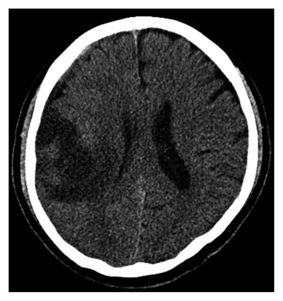
A 76-year-old man presented to another hospital with recurrent left side weakness that had begun several months prior without a history of head trauma. Brain computed tomography (CT) was performed and revealed acute and chronic mixed type SDH on the right convexity (Fig. 1). At that time, the medical team made a decision for the patient to be on conservative care, because the amount of SDH was subtle and the patient’s symptoms were mild.
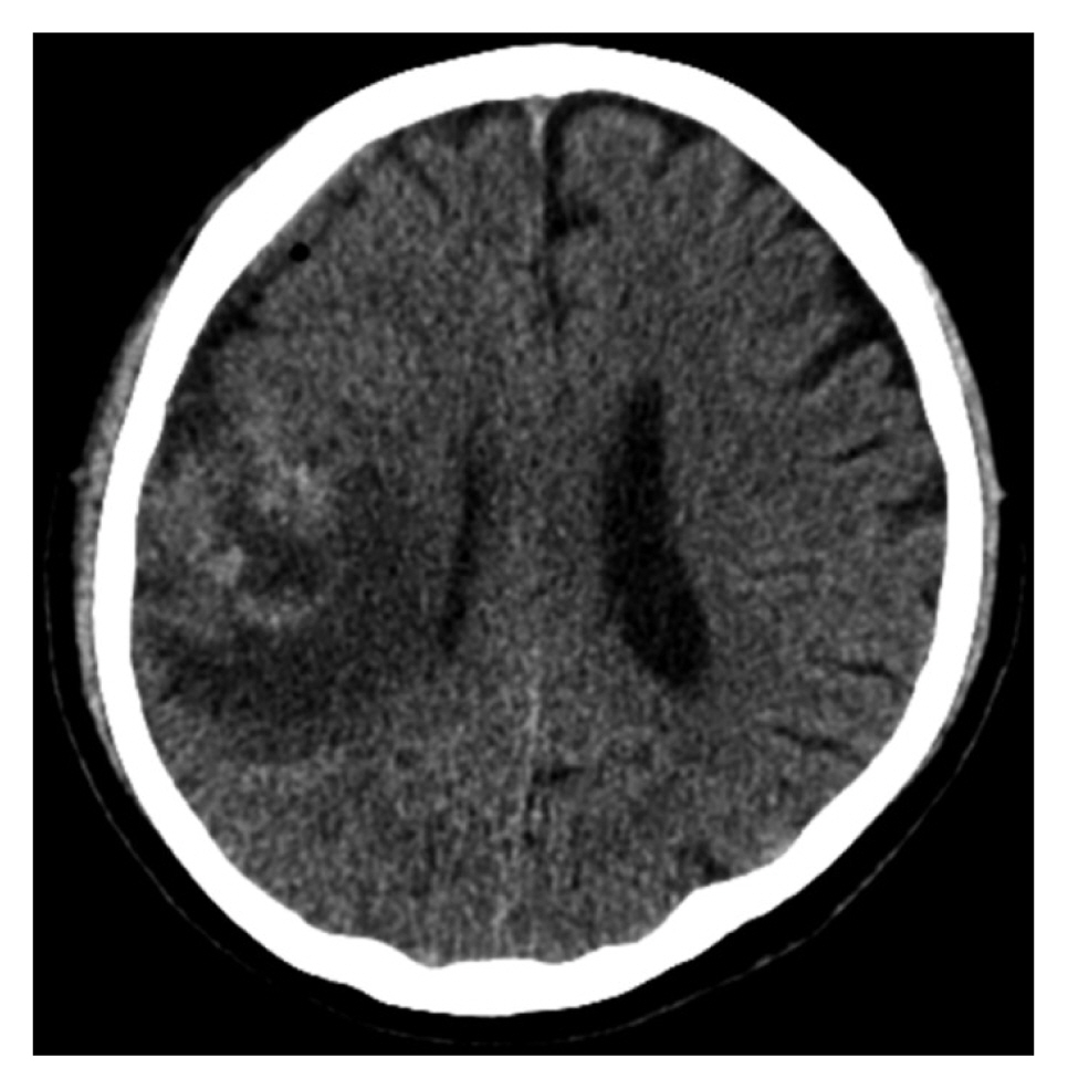
After one month, the patient’s symptoms did not improve and left side weakness and hypoesthesia was aggravated; hence, the patient has transferred our hospital for further workup and management. Brain CT was performed and revealed SDH and new low-density lesion on the right frontoparietal area (Fig. 2). First, we assumed the newly observed lesion was a fluid collection from subdural hygroma or cerebral infarction. We expected the symptoms to be aggravated due to SDH and fluid collection and decided to perform surgical treatment via burr hole trephination for SDH drainage. After surgical management, a dark brownish fluid, a hallmark of typical chronic SDH, was drained. However, the amount of fluid drained was not significant and the patient’s symptoms were not improved.
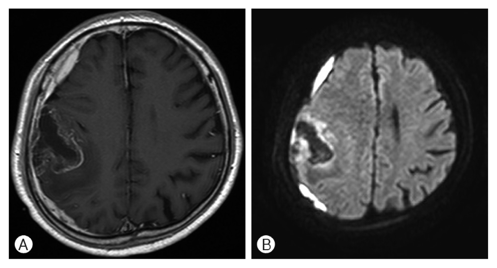
Postoperative brain CT showed a subtle reduction in SDH and a new high-density lesion at the site of the previous low-density lesion (Fig. 3). For evaluation of this new lesion, a full-sequence brain magnetic resonance (MR) imaging (MRI) was performed. Brain MRI revealed a heterogeneous peripheral enhancing irregular mass-like lesion with perilesional edema, for which we could not exclude brain tumor (Fig. 4). Craniotomy was performed for mass-lesion biopsy and removal. The result of mass-lesion biopsy was GBM.
The results of biopsy was GBM
Results of immunohistochemistry:
  1. GFAP: positive in background

  2. p53: strong and diffuse positivity

  3. Ki-67: increased labeling index (about over 15%)

  4. CD34: increased vasculature with meshwork pattern

  5. IDH-1: negative

GFAP: glial fibrillary acidic protein; IDH-1: isocitrate dehydrogenase 1.

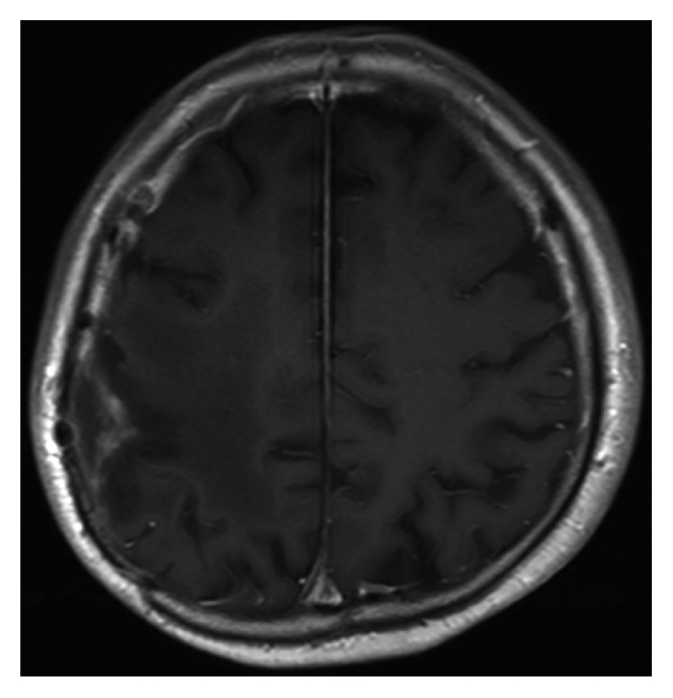
Follow-up brain MRI examination showed the lesion was almost totally removed(Fig. 5). The subsequent concordant radiochemotherapy, which was initiated four weeks after surgery, was tolerated very well.

DISCUSSION

In general, patients with SDH have a history of head trauma. However, some SDHs without trauma history is observed. A broad cause of spontaneous SDH should be considered including vascular and even neoplastic origin. Coombs et al.5) reported that among the 63% of cases where patient sex was specified, there was a 2:1 male predominance; overall, the majority of causes of spontaneous SDH were arterial (61.5%), then idiopathic (10.8%), coagulopathic (10.1%), neoplastic (5.4%), and spontaneous intracranial hypotension (5.4%).
The known vascular origins of non-traumatic SDH are ruptures of small cortical arteries or intracranial aneurysms, rupture of arteriovenous malformation (AVM), and hypertensive or amyloid angiopathic intracerebral hematoma (ICH) perforating the brain surface and arachnoid membrane1,3,5,6,20,22). Aneurysmal rupture usually produces subarachnoid hemorrhage (SAH) with or without ICH and/or intraventricular hematoma (IVH). Aneurysmal SDH with or without SAH is rare, with an incidence estimated to be between 0.5% and 7.9%20). A rare case of non-traumatic acute SDH without SAH, which was caused by rupture of a cortical artery aneurysm, was reported and the patient did not have an infection, inflammation, neoplasm, or head trauma1). The pathogenesis of distal MCA or cortical artery aneurysms includes infectious or neoplastic emboli, arteritis, head trauma, and arterial dissection21). In cases of AVM rupture, ICH is the most common, followed by IVH and SAH. Some authors reported rare cases of SDH due to rupture of cortical AVM and thought AVM vessel adhesion to the arachnoid membrane and strain on the arachnoid membrane could have led to the formation of SDH rather than SAH or ICH3,6). Rupture of a leptomeningeal artery adjacent to the dura, with extravasation into both the brain parenchyma and the subdural space, is a possible explanation of SDH and non-traumatic primary lobar ICH22).
Spontaneous ICH is the most common bleeding complication in a patient with coagulopathy or on antiplatelet or anticoagulant therapy, followed by non-traumatic SDH or infrequent SAH2,7). Although the pathogenesis of non-traumatic SDH with coagulopathy or anticoagulant therapy remains unclear, many studies reported that the mortality rate in patients with acute or chronic SDH and coagulopathy was higher7,13).
Spontaneous intracranial hypotension (SIH) is another well-known cause of non-traumatic SDH. SIH is sometimes associated with bilateral chronic SDH. The incidence of SDH among SIH patients ranges from 16% to 57%, predominantly in males, whereas SIH without SDH predominantly occurs in females8,14,18,30). Low cerebrospinal fluid pressure generates tears in the bridging veins of the dural border cell layer, followed by vein rupture5,25).
Intracranial tumors are a well-known cause of hemorrhage into the brain parenchyma. Clinical and autopsy studies have reported that brain tumors were found in 0.9% to 11% of spontaneous ICH19,26,29). However, non-traumatic SDH associated with neoplasm is believed to occur rarely and has several pathogenesis including hemorrhagic effusion from malignant tissue, dural vessel obstruction by neoplastic cells, angio-demoplastic reaction of the dura to tumor infiltration, and direct hematogenous metastasis of malignant tumor17,23,26,29). The incidence of brain hemorrhage from metastatic and primary intracranial malignant tumors is similar. Lung cancer9), melanoma19), and renal cell cancer28), as well as choriocarcinoma24), are implicated most frequently when brain hemorrhage results from a metastatic lesion. Oligodendroglioma or GBM primary intracranial tumors are often reported to be associated with hemorrhage4,10,11, 15,16,27). One retrospective study reported that the hemorrhage rate for mixed oligodendroglioma/astrocytoma was higher than GBM12).
Most SDHs are generally thought to be caused by head trauma and can happen in any age group. Brain CT scans are usually sufficient for diagnosis in cases of traumatic SDH. When a SDH patient presents without a history of head trauma, the differential diagnosis should include various causes, as mentioned above, and be evaluated via neuroimaging modalities including brain MRI and CT, and/or MR angiography in addition to complete medical history.

CONCLUSION

SDH is typically a sequela of head trauma; however, some cases without a definitive history of head trauma. So-called non-traumatic or spontaneous SDH can occur due to various causes like vascular lesions, neoplasms, coagulopathies, and SIH. When a patient suffers from SDH without a clear history of head trauma, it is wise to conduct additional examinations, such as CT angiography or brain MRI, to help determine the cause of SDH and to plan further treatment.

CONFLICTS OF INTEREST

No potential conflict of interest relevant to this article was reported.

Fig. 1
Brain computed tomography scan shows mixed acute and chronic subdural hematoma in the right frontoparietal area.
nv-7-2-110f1.jpg
Fig. 2
Preoperative brain computed tomography scan reveals subdural hematoma and a low density lesion in the right frontoparietal area.
nv-7-2-110f2.jpg
Fig. 3
Brain computed tomography scan after burr hole craniotomy shows a new high density lesion in the right frontoparietal area.
nv-7-2-110f3.jpg
Fig. 4
(A) Brain magnetic resonance imaging (MRI) (T1-weighted image with enhancement, axial view) shows heterogeneous peripheral enhancing irregular mass in the right frontoparietal lobe. (B) Brain MRI (diffusion-weighted image, axial view) shows low signal intensity.
nv-7-2-110f4.jpg
Fig. 5
Brain magnetic resonance imaging (T1-weighted image with enhancement, axial views) 1-year after surgical tumor resection followup shows stable glioblastoma status at the tumor resection site and no evidence of local tumor recurrence.
nv-7-2-110f5.jpg

REFERENCES

1. Awaji K, Inokuchi R, Ikeda R, Haisa T: Nontraumatic pure acute subdural hematoma caused by a ruptured cortical middle cerebral artery aneurysm: Case report and literature review. NMC Case Rep J 3:63-66, 2016
crossref pmid pmc
2. Butler AC, Tait RC: Management of oral anticoagulant-induced intracranial haemorrhage. Blood Rev 12:35-44, 1998
crossref pmid
3. Choi HJ, Lee JI, Nam KH, Ko JK: Acute spontaneous subdural hematoma due to rupture of a tiny cortical arteriovenous malformation. J Korean Neurosurg Soc 58:547-549, 2015
crossref pmid pmc
4. Chrastina J, Novák Z, Riha I, Ghallab K: Primary brain tumor as a rare cause of acute subdural hematoma. Rozhl Chir 88:549-553, 2009
pmid
5. Coombs JB, Coombs BL, Chin EJ: Acute spontaneous subdural hematoma in a middle-aged adult: case report and review of the literature. J Emerg Med 47:e63-e68, 2014
crossref pmid
6. Datta NN, Chan KY, Kwok JC, Poon CY: Posterior fossa subdural hematoma due to ruptured arteriovenous malformation. Case report. Neurosurg Focus 8:ecp1, 2000

7. Hart RG, Boop BS, Anderson DC: Oral anticoagulants and intracranial hemorrhage. Facts and hypotheses Stroke 26:1471-1477, 1995
crossref pmid
8. Hashizume K, Watanabe K, Kawaguchi M, Fujiwara A, Furuya H: Evaluation on a clinical course of subdural hematoma in patients undergoing epidural blood patch for spontaneous cerebrospinal fluid leak. Clin Neurol Neurosurg 115:1403-1406, 2013
crossref pmid
9. Ishii N, Yokosuka K, Sekihara Y, Hirano K, Suzuki Y, Ishii R: A case of subdural hematoma associated with dural metastasis of lung cancer. No Shinkei Geka 35:511-513, 2007
pmid
10. Kambe A, Hosoya T, Sakamoto M, Kondo S, Kurosaki M: High-grade glioma masquerading as a small cerebral hemorrhage: A case report. Yonago Acta Med 62:305-307, 2019
crossref pmid pmc
11. Kidoguchi M, Isozaki M, Hirose S, Kitai R, Kikuta KI: A case of subcortical intracerebral hemorrhage caused by underlying oligodendroglioma diagnosed through long-term follow-up. No Shinkei Geka 45:233-238, 2017
pmid
12. Kondziolka D, Bernstein M, Resch L, Tator CH, Fleming JF, Vanderlinden RG, et al.: Significance of hemorrhage into brain tumors: clinicopathological study. J Neurosurg 67:852-857, 1987
crossref pmid
13. König SA, Schick U, Döhnert J, Goldammer A, Vitzthum HE: Coagulopathy and outcome in patients with chronic subdural haematoma. Acta Neurol Scand 107:110-116, 2003
crossref pmid
14. Lai TH, Fuh JL, Lirng JF, Tsai PH, Wang SJ: Subdural haematoma in patients with spontaneous intracranial hypotension. Cephalalgia 27:133-138, 2007
crossref pmid
15. Lee J, Kim MS, Kim YZ: Extensive pachymeningeal dissemination of glioblastoma mimicking chronic subdural hematoma: A case report. Brain Tumor Res Treat 7:39-43, 2019
crossref pmid pmc
16. Licata B, Turazzi S: Bleeding cerebral neoplasms with symptomatic hematoma. J Neurosurg Sci 47:201-210, 2003
pmid
17. Minette SE, Kimmel DW: Subdural hematoma in patients with systemic cancer. Mayo Clin Proc 64:637-642, 1989
crossref pmid
18. Mokri B: Low cerebrospinal fluid pressure syndromes. Neurol Clin 22:55-74, vi, 2004
crossref pmid
19. Navi BB, Reichman JS, Berlin D, Reiner AS, Panageas KS, Segal AZ, et al.: Intracerebral and subarachnoid hemorrhage in patients with cancer. Neurology 74:494-501, 2010
crossref pmid pmc
20. Nozar A, Philippe D, Fabrice P, Silvia M, Marc T: Acute pure spontaneous subdural haematoma from ruptured intracranial aneurysms. Interv Neuroradiol 8:393-398, 2002
crossref pmid pmc
21. Nussbaum ES, Madison MT, Goddard JK, Lassig JP, Nussbaum LA: Peripheral intracranial aneurysms: management challenges in 60 consecutive cases. J Neurosurg 110:7-13, 2009
crossref pmid
22. Patel PV, FitzMaurice E, Nandigam RN, Auluck P, Viswanathan A, Goldstein JN, et al.: Association of subdural hematoma with increased mortality in lobar intracerebral hemorrhage. Arch Neurol 66:79-84, 2009
crossref pmid pmc
23. Reichman J, Singer S, Navi B, Reiner A, Panageas K, Gutin PH, et al.: Subdural hematoma in patients with cancer. Neurosurgery 71:74-79, 2012
crossref pmid
24. Rocque BG, Başkaya MK: Spontaneous acute subdural hematoma as an initial presentation of choriocarcinoma: a case report. J Med Case Rep 2:211, 2008
crossref pmid pmc
25. Schievink WI: Spontaneous spinal cerebrospinal fluid leaks and intracranial hypotension. JAMA 295:2286-2296, 2006
crossref pmid
26. Schrader B, Barth H, Lang EW, Buhl R, Hugo HH, Biederer J, et al.: Spontaneous intracranial haematomas caused by neoplasms. Acta Neurochir (Wien) 142:979-985, 2000
crossref pmid
27. Specht CS, Pinto-Lord C, Smith TW, DeGirolami U, Suran E, Marshall PC, et al.: Spontaneous hemorrhage in a mixed glioma of the cerebellum: case report. Neurosurgery 19:278-281, 1986
crossref pmid
28. Spetzger U, Mull M, Sure U, Gilsbach J: Subarachnoid and intraventricular hemorrhage caused by hypernephroma metastasis, accompanied by innocent bilateral posterior communicating artery aneurysms. Surg Neurol 44:275-278, 1995
crossref pmid
29. Wakai S, Yamakawa K, Manaka S, Takakura K: Spontaneous intracranial hemorrhage caused by brain tumor: its incidence and clinical significance. Neurosurgery 10:437-444, 1982
pmid
30. Yoon SH, Chung YS, Yoon BW, Kim JE, Paek SH, Kim DG: Clinical experiences with spontaneous intracranial hypotension: a proposal of a diagnostic approach and treatment. Clin Neurol Neurosurg 113:373-379, 2011
crossref pmid
Editorial Office
Department of Neurosurgery, Dankook University Hospital
201, Manghyang-ro, Dongnam-gu, Cheonan-si, Chungcheongnam-do 31116, South Korea
TEL: +0082-41-550-6280   FAX: +82-41-556-0524   
E-mail: spine1225@naver.com , mousehunt7@naver.com
About |  Browse Articles |  Current Issue |  For Authors and Reviewers
Copyright © The Korean Society of Peripheral Nervous System.                 Developed in M2PI