| Home | E-Submission | Sitemap | Contact Us |  
top_img
The Nerve > Volume 10(1); 2024 > Article
Song, Lee, Ryu, Lee, Suh, Lee, and Cho: Glioblastoma Presenting with the Radiological Features of a Meningioma: A Case Report and Literature Review

Abstract

Glioblastomas, which are the most common primary malignant brain tumors in adults, occasionally exhibit radiological features similar to those of other diseases. However, instances of them mimicking meningiomas are rare. This article describes a rare case of a glioblastoma presenting with radiological features similar to those of a meningioma. A 55-year-old man was admitted following 2 months of gait disturbance, mild dysarthria, and dizziness. Magnetic resonance imaging suggested an extra-axial tumor in the interhemispheric fissure exhibiting a cerebrospinal fluid cleft sign, which was indicative of a meningioma. Bifrontal craniotomy for tumor resection revealed a highly vascularized, yellowish-gray friable mass within the interhemispheric fissure. The pathological diagnosis was glioblastoma. The patient’s symptoms improved following treatment with whole-brain radiotherapy and temozolomide chemotherapy, and his condition was stable during a 5-month follow-up period. Glioblastomas may present with radiological features similar to those of meningiomas, making it challenging for physicians to establish an accurate diagnosis and effective treatment plan.

INTRODUCTION

Glioblastoma, which is predominantly an intra-axial neoplasm, is the most common primary malignant brain tumor4). Glioblastoma may exhibit characteristic radiological features of other diseases, such as arteriovenous malformations, viral encephalitis, lymphomas, subacute strokes, and cerebral abscesses, upon isointensity (computed tomography [CT]) or magnetic resonance imaging (MRI). However, glioblastomas mimicking the radiological features of a meningioma are rare. Herein, we describe a patient with glioblastoma exhibiting the radiological features of a meningioma.

CASE REPORT

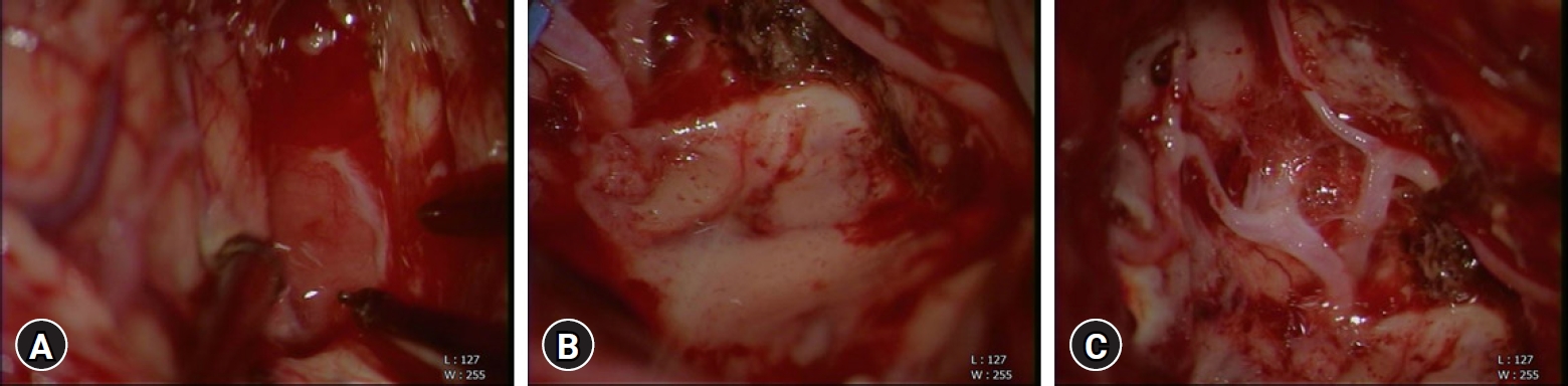
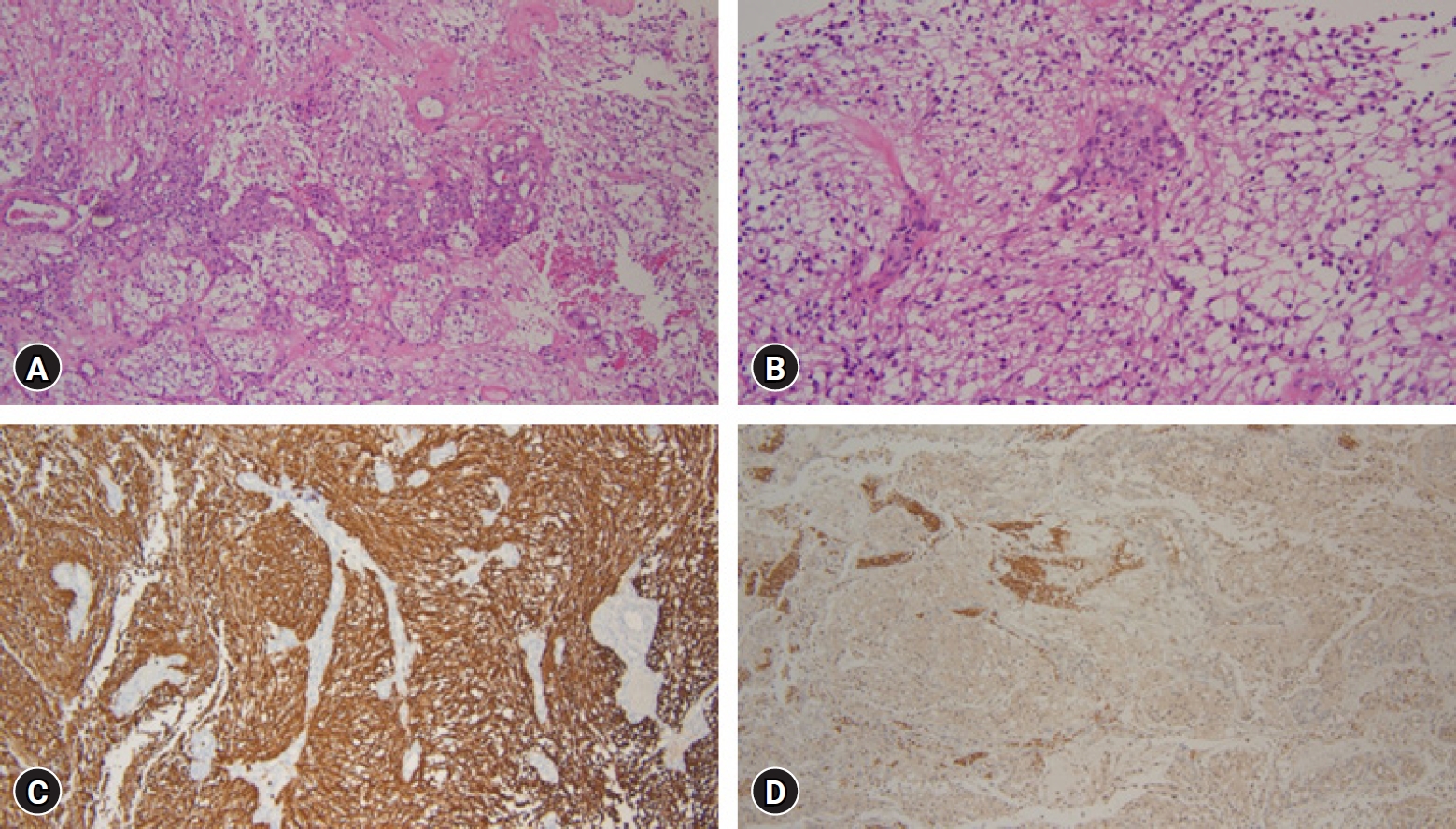
A 55-year-old male patient presented with gait disturbance, mild dysarthria, and dizziness over two months. CT imaging revealed ventriculomegaly and a discernible mass in the interhemispheric fissure (Fig. 1A). Subsequent MRI scans identified a heterogeneously enhancing extra-axial mass, measuring 2.3×2.3×2.5 cm, with well-defined margins (Fig. 1B). Isointensity to the gray matter in the T1-weighted sequence (Fig. 1C) and slight hyperintensity relative to the gray matter in the T2-weighted sequence (Fig. 1D) were observed. Notably, the T2-weighted sequence demonstrated a cerebrospinal fluid (CSF) cleft (Fig. 1D) between the tumor and adjacent brain cortex, leading to an initial radiological diagnosis of meningioma. Two days later, the patient underwent a bifrontal craniotomy for tumor resection, which revealed a highly vascularized, yellowish-gray, friable mass within the interhemispheric fissure (Fig. 2). Macroscopic examination revealed no significant infiltration of the tumor into the brain parenchyma, and subtotal mass removal was performed. Intraoperative pathological assessment suggested a high-grade glioma. Histopathological examination of the surgical specimen revealed atypical glial cells and marked endothelial and microvascular proliferation, but no necrotic changes (Fig. 3A, B). Immunohistochemical analysis demonstrated diffuse positivity for S-100, phosphatase and tensin homolog, and glial fibrillary acidic protein in the tumor cells (Fig. 3C). In contrast, the tumor cells were negative for epithelial membrane antigen, aiding in the differentiation of the specimen from a meningioma. The proliferation index, as indicated by Ki-67, was intermediate, ranging between 16% and 30%. Molecular investigations revealed no O-6-methylguanine-DNA methyltransferase gene methylation or isocitrate dehydrogenase (IDH) 1 mutations (Fig. 3D). These findings were consistent with those of glioblastoma, IDH wild-type, and World Health Organization (WHO) grade IV. Postoperative MRI, conducted one day after surgery revealed no residual enhanced lesions (Fig. 4). Subsequently, the patient underwent whole-brain radiotherapy and chemotherapy with temozolomide, and was discharged without any neurological deficits. The patient remained symptom-free for approximately five months after surgery.

DISCUSSION

Definitive diagnosis of an intracranial mass depends on pathological and molecular analyses. However, radiological characteristics serve as the primary means of gauging the nature of the tumor prior to performing a biopsy. Determining whether intracranial masses are intra- or extra-axial is very important as this affects the diagnosis, treatment plan, and prognosis. Extra-axial masses include meningiomas, schwannomas, metastatic lesions, arachnoid cysts, epidermoids, dermoids, chordomas, and eosinophilic granulomas5,15). The characteristic radiological features differentiating extra-axial from intra-axial masses include local bone changes, white matter buckling (inward compression of the gray-white junction), pseudocapsule (signal void of the dura or displaced vessels), CSF cleft (CSF or cortical vessels entrapped between the tumor and underlying cortex), and dural tail sign (enhancement of the dura infiltrating away from the lesion)2,5,6,9,15).
Glioblastomas typically present as invasive intra-axial masses with irregular margins. The typical characteristics of glioblastomas are hypodense signal change on the T1-weighted sequence and hyperintense signal change on the T2-weighted sequence. On contrast-enhanced T1-weighted MRI scans, glioblastomas classically appear hypointense to isointense, displaying a ring-enhancing pattern, often accompanied by vasogenic edema, central necrosis, and infiltration into the white matter nerve bundles1). Conversely, meningiomas generally appear as lobular, extra-axial masses with well-defined margins, exhibiting avid, homogeneous enhancement on contrast-enhanced T1-weighted MRI scans, although they may occasionally present with areas of central necrosis or calcification that do not enhance14,18). The typical characteristics of meningiomas are iso to slight hypodense signal change on the T1-weighted sequence and iso to hyperintense signal change on the T2-weighted sequence. Distinct radiological features, such as the dural tail sign, CSF cleft sign, sunburst or spoke-wheel appearance of vessels, and either osteolysis or hyperostosis, are indicative of meningiomas13,18).
In the present case, the patient's MRI findings— a lobular, extra-axial mass with a well-circumscribed margin, and CSF cleft sign—were consistent with a meningioma than a glioblastoma. Therefore, even if the radiological findings are consistent with those of a meningioma, the possibility of a glioblastoma should not be excluded.
The WHO 2016 Classification of Tumors of the Central Nervous System (WHO CNS4) categorizes astrocytic tumors into astrocytomas, either IDH mutant or wild type, and glioblastomas, also IDH mutant or wild type. However, the WHO 2021 Classification of Tumors of the Central Nervous System (WHO CNS5) reclassified astrocytic gliomas into astrocytoma, IDH-mutant, and glioblastoma, IDH-wild type. IDH-mutant astrocytomas are categorized as CNS WHO grades 2, 3, or 4 in the WHO CNS5, whereas IDH-wild type astrocytomas, synonymous with the diagnosis of glioblastoma, are classified as grade 4. The WHO CNS4 classification of glioblastoma, IDH-mutant (secondary glioblastoma), and IDH-wild type (primary glioblastoma) was updated to separate entities: astrocytoma, IDH-mutant, and glioblastoma, according to the WHO CNS5. Table 1 summarizes the differences between the WHO CNS4 and CNS510).
To our knowledge, few cases of glioblastoma-mimicking meningioma have been reported. As mentioned above, the diagnostic criteria for glioblastomas have changed. However, reports published in the literature can still be used as a reference. Dabboucy et al.3) reported a case of primary extra-axial glioblastoma in the left temporal area that exhibited a dural tail and CSF cleft sign. Patel et al.14) described two cases of glioblastoma mimicking meningioma. The first case involved a heterogeneously enhancing right temporoparietal mass with extensive contact along the right tentorium, a CSF cleft sign, and a dural tail sign, while the second case involved a left parasagittal, heterogeneously enhancing mass adjacent to the falx, and featured a dural tail sign. Taghipour Zahir et al.16) reported a glioblastoma with calvarial involvement. Lee et al.12) described a case of a glioblastoma within the cerebellopontine angle, while Karthigeyan et al.7) reported a case of glioblastoma within a petroclival location. Kayaci et al.8) and Varma et al.17) described cases of glioblastomas in the mid one-third falx. Table 2 summarizes the reported cases of glioblastomas mimicking meningiomas.
Magnetic resonance spectroscopy (MRS) can be used to distinguish between benign and malignant tumors, thus assisting in diagnosis. In a study by Lee et al.12), MRS was utilized for cerebellopontine angle tumors that rapidly increased in size over three months. The findings demonstrated increased choline-to-creatinine ratios and lactate levels, whereas N-acetyl aspartate levels were decreased, suggesting malignancy. Similarly, Karthigeyan et al.7) conducted MRS on a petroclival tumor in a 27-year-old patient with a relatively short disease duration and rapidly progressing symptoms. Although the patient's CT scan showed calcification, MRS revealed a choline-to-creatinine ratio of 2.5, indicating malignancy11). In cases exhibiting symptoms suggestive of malignancy, as in the two cases mentioned above, differentiating the tumors using MRS appears beneficial, even if the radiological images suggest a benign nature. However, when symptoms and imaging findings lean towards benign indications, the use of MRS may require further contemplation, although it undoubtedly contributes to making the diagnosis.

CONCLUSION

Glioblastomas may present with radiological features similar to those of meningiomas, making it precise diagnoses and formulating treatment plans challenging for physicians. Additional diagnostic tests, such as MRS, can be instrumental in aiding the diagnosis; however, their clinical indications require further deliberation.

CONFLICTS OF INTEREST

No potential conflict of interest relevant to this article was reported.

Fig. 1.
(A) Axial computed tomography image demonstrates mass on interhemispheric fissure with hypodensity. (B) Post-contrast axial T1-weighted sequence demonstrates heterogenous. (C) Axial T1-weighted sequence demonstrates isointensity relative to grey matter. (D) Axial T2-weighted sequence demonstrates slight hyperintensity relative to grey matter with a prominent surrounding cerebrospinal fluid signal intensity cleft between the mass and the adjacent frontal cortex.
nerve-2024-00507f1.jpg
Fig. 2.
(A, B) Highly vascularized, yellowish-gray friable mass within the interhemispheric fissure was identified in operation field. (C) After resection, adjacent vessels were visible.
nerve-2024-00507f2.jpg
Fig. 3.
(A) Histopathological examination of the surgical specimen showed atypical glial cells, marked endothelial proliferation (hematoxylin and eosin [H&E], ×100 magnification). (B) Marked microvascular proliferation in the tumor (H&E, ×200 magnification). (C) Immunohistochemical staining for glial fibrillary acidic protein reveals diffuse immunoreactivity in the tumor cells (×100 magnification). (D) Immunohistochemical staining for isocitrate dehydrogenase 1 reveals negative results in the tumor cells (×100 magnification).
nerve-2024-00507f3.jpg
Fig. 4.
Postoperative magnetic resonance imaging showed nearly total removal of mass and no definite enhance lesion.
nerve-2024-00507f4.jpg
Table 1.
Differences between the WHO CNS4 and CNS5
The WHO 2016 Classification of Tumors of the CNS (WHO CNS4) The WHO 2021 Classification of Tumors of the CNS (WHO CNS5)
Diffuse astrocytoma, IDH-mutant, WHO grade II Astrocytoma, IDH-mutant, CNS WHO grade 2
Anaplastic astrocytoma, IDH-mutant, WHO grade III Astrocytoma, IDH-mutant, CNS WHO grade 3
Glioblastoma, IDH-mutant WHO grade IV (secondary glioblastoma) Astrocytoma, IDH-mutant, CNS WHO grade 4
Diffuse astrocytoma, IDH-wildtype, WHO grade II Glioblastoma, IDH-wildtype, CNS WHO grade 4
Anaplastic astrocytoma, IDH-wildtype, WHO grade III
Glioblastoma, IDH-wildtype, WHO grade IV (primary glioblastoma)

WHO: World Health Organization; CNS: central nervous system; IDH: isocitrate dehydrogenase.

Table 2.
Previous case reports that have described glioblastomas mimicking meningiomas
References Year Age/Sex Location Magnetic resonance imaging Additional study Treatment Prognosis
Dabboucy et al.3) 2021 76/F Left temporal T2, hyperintense, w/ CSF cleft sign; Heterogenous enhancement mass w/ broad dural contact w/ DTS None Gross total resection followed by whole brain RT & TMZ Alive after 6 months
Patel et al.14) 2016 57/F Right temporo-parietal T2, hyperintense, w/ vasogenic edema and CSF cleft sign; heterogenous enhancement mass w/ broad dural contact w/ DTS Cerebral angiography & embolization Total resection followed by whole brain RT & TMZ Not available
60/M Left parasagittal Heterogenous enhancement mass w/ a DTS Cerebral angiography & embolization Resection followed by whole brain RT & TMZ Recurrence after 22 months, dead after 28 months
Taghipour Zahir et al.16) 2018 60/M Right frontal calvarium Heterogenous enhancement mass w/ infiltration of bone, dura, and galea None Biopsy followed by Whole brain RT & TMZ Dead after 7 months
Lee et al.12) 2017 71/F Left cerebellopontine angle T2, hyperintense; peripheral enhancement mass w/ central necrosis MRS Biopsy followed by Whole brain RT & TMZ Alive after
1 year
Karthigeyan et al.7) 2017 27/F Left petroclival T1, hypointense; T2, hyperintense; heterogeneous enhancement mass w/ DTS MRS Subtotal resection followed by RT Not available
Kayaci et al.8) 2014 19/M Middle one-third falx T2, iso-hyperintense; heterogeneous enhancement mass w/ edema None Subtotal resection followed by RT & TMZ Alive after 5 months
Varma et al.17) 2020 30/F Middle one-third falx T1, heterogenous; T2, heterogenous w/ buckling of the cortex; heterogeneous enhancement mass w/ edema None Resection Not available
Present case 2024 55/M Interhemispheric fissure T1, isointense; T2, hyperintense; heterogeneous enhancement mass w/ CSF cleft sign None Gross total resection followed by whole brain RT & TMZ Alive after 5 months

M: male; F: female; CSF: cerebrospinal fluid; DTS: dural tail sign; RT: radiation therapy; TMZ: temozolomide; MRS: magnetic resonance spectroscopy.

REFERENCES

1. Abd-Elghany AA, Naji AA, Alonazi B, Aldosary H, Alsufayan MA, Alnasser M, et al.: Radiological characteristics of glioblastoma multiforme using CT and MRI examination. J Radiat Res Appl Sci 12:289-293, 2019
crossref
2. Curnes JT: MR imaging of peripheral intracranial neoplasms: extraaxial vs intraaxial masses. J Comput Assist Tomogr 11:932-937, 1987
crossref pmid
3. Dabboucy B, Younes P, Rahbani A, Fahed E, Abadjian G: Primary extra-axial glioblastoma: case report and literature review. Arq Bras Neurocir 40:e368-e373, 2021
crossref
4. Di Carlo DT, Cagnazzo F, Benedetto N, Morganti R, Perrini P: Multiple high-grade gliomas: epidemiology, management, and outcome. A systematic review and meta-analysis. Neurosurg Rev 42:263-275, 2019
crossref pmid pdf
5. Drevelegas A: Extra-axial brain tumors. Eur Radiol 15:453-467, 2005
crossref pmid pdf
6. George AE, Russell EJ: White matter buckling: CT sign of extraaxial intracranial mass. AJR Am J Roentgenol 135:1031-1036, 1980
crossref pmid
7. Karthigeyan M, Ahuja CK, Chatterjee D, Salunke P: Radiologic dilemma in an extra-axial petroclival lesion: low grade versus high grade. World Neurosurg 100:713.e1-713.e3, 2017
crossref pmid
8. Kayaci S, Şengöz A, Köksal V, Gunver F, Kiliç K: Glioblastoma multiforme mimicking falx meningioma with achondroplasia. Neurosurg Q 24:53-55, 2014
crossref
9. Kim J, Hong CK, Suh SH, Jung WS, Ahn SJ: The added value of the CSF cleft on ADC in distinguishing extra-axial from intra-axial tumors. Clin Imaging 45:81-85, 2017
crossref pmid
10. Kurokawa R, Kurokawa M, Baba A, Ota Y, Pinarbasi E, Camelo-Piragua S, et al.: Major changes in 2021 world health organization classification of central nervous system tumors. Radiographics 42:1474-1493, 2022
crossref pmid
11. Laino ME, Young R, Beal K, Haque S, Mazaheri Y, Corrias G, et al.: Magnetic resonance spectroscopic imaging in gliomas: clinical diagnosis and radiotherapy planning. BJR Open 2:20190026, 2020
crossref pmid pmc
12. Lee JH, Kim JH, Kwon TH: Primary glioblastoma of the cerebellopontine angle: case report and review of the literature. J Korean Neurosurg Soc 60:380-384, 2017
crossref pmid pmc pdf
13. Lyndon D, Lansley JA, Evanson J, Krishnan AS: Dural masses: meningiomas and their mimics. Insights Imaging 10:11, 2019
crossref pmid pmc pdf
14. Patel M, Nguyen HS, Doan N, Gelsomino M, Shabani S, Mueller W: Glioblastoma mimicking meningioma: report of 2 cases. World Neurosurg 95:624.e9-624.e13, 2016
crossref pmid
15. Politi M, Alektorov K, Moulopoulos LA, Papanagiotou P: Extra-axial brain tumours and cysts. Hell J Radiol 5:30-42, 2020

16. Taghipour Zahir S, Mortaz M, Baghi Yazdi M, Sefidrokh Sharahjin N, Shabani M: Calvarium mass as the first presentation of glioblastoma multiforme: A very rare manifestation of high-grade glioma. Neurochirurgie 64:76-78, 2018
crossref pmid
17. Varma A, Gopalakrishnan CV, Kachare N, Panikar D: Glioblastoma masquerading as a parafalcine meningioma: a pathological surprise! J Neurosci Rural Pract 11:668-669, 2020
crossref pmid pmc
18. Watts J, Box G, Galvin A, Brotchie P, Trost N, Sutherland T: Magnetic resonance imaging of meningiomas: a pictorial review. Insights Imaging 5:113-122, 2014
crossref pmid pmc pdf
Editorial Office
Department of Neurosurgery, Dankook University Hospital
201, Manghyang-ro, Dongnam-gu, Cheonan-si, Chungcheongnam-do 31116, South Korea
TEL: +0082-41-550-6280   FAX: +82-41-556-0524   
E-mail: spine1225@naver.com , mousehunt7@naver.com
About |  Browse Articles |  Current Issue |  For Authors and Reviewers
Copyright © The Korean Society of Peripheral Nervous System.                 Developed in M2PI