| Home | E-Submission | Sitemap | Contact Us |  
top_img
The Nerve > Volume 10(1); 2024 > Article
Jang, Sheen, Han, Lee, Choi, Bock, and Sohn: Postoperative Thoracic Cord Compression Induced by a Dural Sealant System (DuraSeal®): A Case Report and Literature Review

Abstract

Cerebrospinal fluid (CSF) leakage is a rare complication that can occur due to dural defects during spinal surgery, hindering the improvement of the surgical site and increasing the possibility of infection. DuraSeal® is a dural sealing adhesive that prevents CSF leakage and is used as an adjunct to enable the watertight repair of dural defects when the dura is damaged during spinal surgery. In the present case, DuraSeal® was applied to repair a dural defect in the surgical area after thoracic spine surgery, and no neurological problems occurred immediately after surgery. However, a day later, the patient’s paraparesis worsened; therefore, reoperation was performed and the symptoms improved.

INTRODUCTION

Cerebrospinal fluid (CSF) leakage is a rare complication that can occur in spinal surgery. Postoperative CSF leakage can induce delayed healing, wound infections, and intracranial hypotension, which causes neurological symptoms such as postural headache, vomiting, and dizziness3,5,11-13). Repair of CSF leakage due to dural defects that may occur during surgery is very important and Dural Sealant System can be used in this case. DuraSeal® (Integra LifeSciences, Princeton NJ, USA) is used to repair dural defects incurred during surgery and is a proven product approved by the U.S. Food and Drug Administration (FDA) in 20051,5). Several cases of spinal cord compression caused by DuraSeal® have been reported. We report a rare case of DuraSeal® causing cord compression during thoracic surgery due to dural defects. This report further supports that DuraSeal® may compress the spinal cord and lead to neurological symptoms.

CASE REPORT

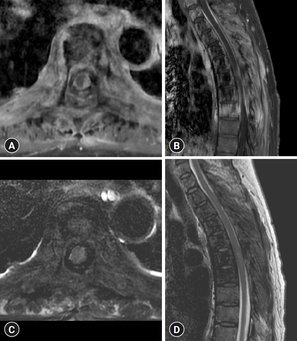
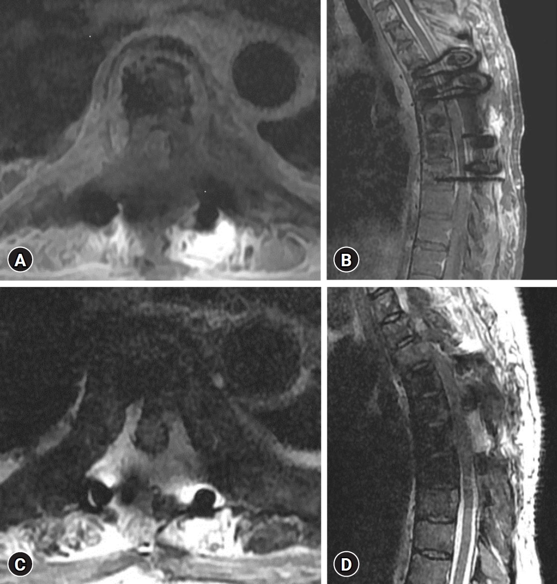
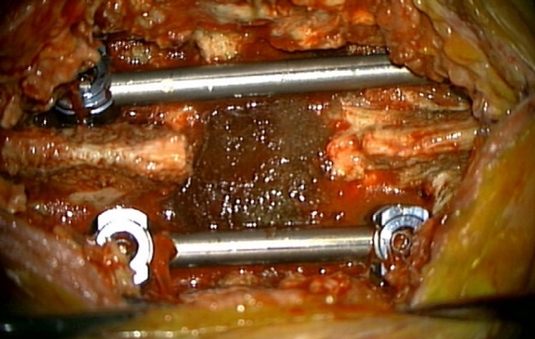
A 57-year-old female patient who was hospitalized in the hematology-oncology department due to breast cancer was transferred to the neurosurgery department for treatment after spinal metastasis was confirmed on magnetic resonance image (MRI) performed due to lower motor weakness. On physical examination, she had both lower extremity weakness (Medical Research Council [MRC] grading 4/5) and gait disturbance. MRI showed overall contrast enhancement in the range from T1 to T8 level, including the paravertebral, vertebrae body, epidural, and spinous processes, resulting in central canal stenosis, and especially cord compression due to the epidural mass effect at the T6 level (Fig. 1). The tumor removal through T6 laminectomy and T4 to T8 screw fixation was performed. The tumor was totally removed, but there was severe adhesion to the dura and there was a tear in the dura bilaterally. Dural sutures were made to repair the torn dura, and DuraSeal® was applied after sealing the dura surface with TachoComb, a patch-type hemostatic agent. The histopathologic finding was metastatic carcinoma. One day after surgery, the patient's both lower extremity weakness worsened (MRC grading 1/5). An MRI was performed immediately, and it was confirmed that an epidural mass showing a high signal on the T2-weighted image was compressing the spinal cord at the tumor removal site (Fig. 2). The patient underwent emergency wound exploration and decompression. When the wound was opened, the DuraSeal® was exposed and the cord was compressed severely (Fig. 3). DuraSeal® was removed totally, and no hematoma or CSF was seen (Fig. 4). One day after the second surgery, the patient showed improvement in both lower extremity weakness (MRC grading 2+/5).

DISCUSSION

As mentioned earlier, CSF leakage is one of the rare complications that can occur after spinal surgery5). The healing period of surgical areas such as muscles, skin, and bones may be prolonged due to CSF leakage. This can potentially lead to complications such as meningitis, nausea, vomiting, vertigo, tinnitus, postural headache, and fistula formation3,5,13). Therefore, preventing CSF leakage is very important. There are two main ways to repair CSF leakage. It is a direct repair method that repairs dural defects using an absorbable suture line and grafting fat, muscle, and fascia, or an alternative method is repaired by applying a material such as DuraSeal® to the dural defects3,13).
DuraSeal® has been approved as a synthetic sealant by the FDA and is used to promote watertight repair of dural defects that occur after neurosurgery4,5). DuraSeal® is a nontoxic bioabsorbable compound based on polyethylene glycol (PEG) and trilysine, a small amino acid. When PEG is mixed with trilysine, the substances undergo cross-linking almost instantly, resulting in the formation of a flexible layer that adheres to surrounding tissues. it also is broken down through hydrolysis over a period of 4 to 8 weeks, and the resulting byproducts are eliminated through renal clearance. Additionally, the MRI imaging characteristics of DuraSeal® are difficult to distinguish from those of CSF3,5,7,8,10).
As a characteristic of DuraSeal® shown in our case, it applied after surgery can swell by up to 50%, reaching peak expansion within 3 to 14 days, and it can last for almost four weeks3,5,9,13). Due to these characteristics, DuraSeal® may cause worsening of neurological symptoms such as postoperative quadriparesis, quadriplegia, and cauda equina syndrome2,5,6,13). We found cases of spinal cord compression after DuraSeal® application during spinal surgery reported since 2009. These are summarized in Table 1.
In the case summarized in Table 1, DuraSeal® was applied to a dura tear that occurred during surgery, and motor weakness and cauda equina syndrome subsequently occurred. The above symptoms had a latency period from immediately after surgery to 9 days, which was consistent with the peak expansion period of DuraSeal®, which was 3 to 14 days. Reoperation was performed for this, and after confirming expansion of the DuraSeal®, it was removed, and most patients' symptoms improved. Based on this, it appears that it can be inferred that cord compression occurred through DuraSeal® expansion. Additionally, in three of the reviewed cases, the hematoma at the surgical site was not properly drained due to DuraSeal® expansion. This suggests that the combination of hematoma and DuraSeal® may further compress the spinal cord1,4,13).
However, a limitation is that the absolute number of cases of side effects caused by DuraSeal® is small, and additional evaluation appears to be necessary to determine whether it is a direct effect of DuraSeal®. In the author's opinion, cord compression by DuraSeal® is likely to occur in areas where the spinal canal is limited, such as the C-spine or T-spine. However, as symptoms were reported in the L-spine as shown in the table, the cord compression by DuraSeal® is likely to occur to some extent for each level. Additional evaluation appears to be necessary regarding the effect on compression and the amount of DuraSeal® used by the surgeon.

CONCLUSION

In our case, we reported that a dura tear that occurred during surgery was repaired by applying DuraSeal®, but cord compression occurred due to the swelling characteristics of the DuraSeal®, causing neurological symptoms. DuraSeal® is a very useful material for dural repair, but it needs to be used with caution, as in the case reports that have been reported so far and in our case. In particular, care must be taken when using it in spaces where the spinal canal is relatively limited, such as the C-spine and T-spine, rather than the L-spine. When using it, the appropriate amount to fit the limited space must be applied thinly. As a result, it is necessary for the surgeon to recognize the postoperative expansion of the DuraSeal® and prevent complications that may arise from it.

Acknowledgments

This work was supported by a grant of Basic Science Research Program through the National Research Foundation of Korea (NRF) funded by the Ministry of Science, ICT and Future Planning (RS-2023-00209591).

CONFLICTS OF INTEREST

No potential conflict of interest relevant to this article was reported.

Fig. 1.
Preoperative contrast-enhanced T1-weighted magnetic resonance images (MRI), axial (A) and sagittal (B), show an epidural well-enhanced mass at the T6 level compressing the cord from the left side. Preoperative T2-weighted MRI, axial (C) and sagittal (D), show no high signal intensity lesions around the cord other than the mass effect of the spinal cord.
nerve-2024-00535f1.jpg
Fig. 2.
Postoperative contrast-enhanced T1-weighted magnetic resonance images (MRI), axial (A) and sagittal (B), show that an epidural-enhanced mass of the spinal cord has been totally removed. Postoperative T2-weighted MRI, axial (C) and sagittal (D), show an extradural high signal intensity lesion surrounding the cord and compressing it at the level of tumor removal.
nerve-2024-00535f2.jpg
Fig. 3.
Microscope photography of swollen DuraSeal® exposed during reoperation.
nerve-2024-00535f3.jpg
Fig. 4.
Microscope photography of a decompressed spinal cord shown after removal of the swollen DuraSeal® exposed during reoperation.
nerve-2024-00535f4.jpg
Table 1.
Summary of case reports of neurological complications resulting from the use of DuraSeal® during surgery
References Operative site Procedure Postoperative symptoms Latency period Treatment Outcome after reoperation Mechanism
Mulder et al. (2009)7) L4-5 Laminotomy and discectomy Cauda equina syndrome 9 days Revision operation Resolution of cauda equina syndrome, but left calf discomfort and left foot drop DuraSeal® expansion
Thavarajah et al. (2010)11) C5-6 Anterior cervical discectomy and fusion Right-side weakness (upper motor grade 3, lower motor grade 2) 3 hours Exploration and decompression Improved right side motor function (upper grade 4, lower grade 3) DuraSeal® expansion
Lee et al. (2010)4) C3-6 Laminectomy Neck pain with radiation down to hands and fingers bilaterally, sensory change and motor weakness 2 days Exploration and decompression Improved limb strength, but poor finger dexterity bilaterally DuraSeal® expansion with remaining hematoma
Neuman et al. (2012)8) L4-5 Transforaminal lumbar interbody fusion Cauda equina syndrome 2 days Exploration and decompression Resolution of cauda equina syndrome DuraSeal® expansion
Lee et al. (2013)5) C7 Tumor removal Left side weakness (motor grade 1) 8 hours Exploration and decompression Improved motor function (motor grade 2) DuraSeal® expansion
Rustagi and Lavelle (2014)9) C5-6 Circumferential fusion Upper and lower extremity weakness Not reported The family declined further surgery Death 8 weeks after surgery DuraSeal® expansion
Arrotegui (2019)1) C6 Anterior cervical discectomy and fusion Paraparesis of upper and left lower limb (motor grade 2) Urgent Revision operation Slight neurological improvement DuraSeal® expansion with remaining hematoma
Han et al. (2020)3) L1 Laminectomy Left lower extremity weakness (motor grade 1-2) and paresthesia 1 day Exploration and decompression Severe paresthesia resolved and motor function improved (motor grade 3) DuraSeal® expansion
Epstein and Esq. (2021)2) C3-6 Corpectomy and fusion quadriplegia Within 9 hours Revision operation Permanent neurological injury DuraSeal® expansion
Yeh et al. (2022)13) L2-4 Laminectomy and foraminal decompression Cauda equina syndrome 2 days Exploration and decompression Permanent neurological injury DuraSeal® expansion with remaining hematoma

REFERENCES

1. Arrotegui I: Motor injury by compression of spinal cord at cervical level during surgery. J Spine Res Surg 1:23-28, 2019
crossref
2. Epstein NE, Esq JL: Medicolegal corner (spine): Contraindicated use of DuraSeal in anterior cervical spine led to quadriplegia. Surg Neurol Int 12:532, 2021
crossref pmid pmc
3. Han HJ, Jeong JH, Kim JW, Seung WB: Postoperative thecal sac compression induced by hydrogel dural sealant after spinal schwannoma removal. Korean J Neurotrauma 16:99-104, 2020
crossref pmid pmc pdf
4. Lee G, Lee CK, Bynevelt M: DuraSeal-hematoma: concealed hematoma causing spinal cord compression. Spine (Phila Pa 1976) 35:E1522-E1524, 2010
crossref pmid
5. Lee SH, Park CW, Lee SG, Kim WK: Postoperative cervical cord compression induced by hydrogel dural sealant (DuraSeal®). Korean J Spine 10:44-46, 2013
crossref pmid pmc
6. Mughal N, Mishra D, Srivastava A, Naaz N: Gelfoam induced cauda equina syndrome: A case report with review of literature. Glob J Res Anal 9:42-44, 2020
crossref
7. Mulder M, Crosier J, Dunn R: Cauda equina compression by hydrogel dural sealant after a laminotomy and discectomy: case report. Spine (Phila Pa 1976) 34:E144-E148, 2009
crossref pmid
8. Neuman BJ, Radcliff K, Rihn J: Cauda equina syndrome after a TLIF resulting from postoperative expansion of a hydrogel dural sealant. Clin Orthop Relat Res 470:1640-1645, 2012
crossref pmid pmc
9. Rustagi T, Lavelle WF: Spontaneous resolution of cervical cord compression induced by hydrogel (Duraseal). Spine J 14:2511-2512, 2014
crossref pmid
10. Tarapore PE, Mukherjee P, Mummaneni PV, Ames CP: The appearance of dural sealants under MR imaging. AJNR Am J Neuroradiol 33:1530-1533, 2012
crossref pmid pmc
11. Thavarajah D, De Lacy P, Hussain R, Redfern RM: Postoperative cervical cord compression induced by hydrogel (DuraSeal): a possible complication. Spine (Phila Pa 1976) 35:E25-E26, 2010
crossref pmid
12. Xiao Y, Wang B, Chen Y, Chen L, Lou Z, Gong Z: Risk factors of postoperative cerebrospinal fluid leak after craniovertebral junction anomalies surgery: A case-control study. Neurospine 20:255-264, 2023
crossref pmid pmc pdf
13. Yeh KL, Wu SH, Fuh CS, Huang YH, Chen CS, Wu SS: Cauda equina syndrome caused by the application of DuraSeal(TM) in a microlaminectomy surgery: A case report. World J Clin Cases 10:11178-11184, 2022
crossref pmid pmc
Editorial Office
Department of Neurosurgery, Dankook University Hospital
201, Manghyang-ro, Dongnam-gu, Cheonan-si, Chungcheongnam-do 31116, South Korea
TEL: +0082-41-550-6280   FAX: +82-41-556-0524   
E-mail: spine1225@naver.com , mousehunt7@naver.com
About |  Browse Articles |  Current Issue |  For Authors and Reviewers
Copyright © The Korean Society of Peripheral Nervous System.                 Developed in M2PI