INTRODUCTION
Acute subdural hematoma (SDH) is commonly caused by traumatic event that tears the bridging veins in the subdural space6). Otherwise, some spontaneous SDH cases have been reported to be related with grossly anatomical abnormality such as aneurysm and arteriovenous malformation (AVM). Some other cases are associated medical conditions such as coagulopathy, alcohol overuses, and so on1,9).
Spontaneous acute SDH from arterial bleeding has been rarely reported. Munro first described a patient who had acute SDH without a history of head trauma in 1934. Talalla and McKissock introduced eight cases of acute spontaneous SDH based on autopsy finding of spontaneous rupture of cortical artery. After using computed tomography (CT), many authors have reported cases of spontaneous SDH with cortical artery origin that are confirmed by operative finding1,7,9). Some cases have revealed a leakage of cortical artery via cerebral angiography4).
Here, we report a rare case of spontaneous acute SDH of cortical artery origin confirmed by operative finding.
CASE REPORT
A 43-year-old woman was hospitalized for surgical removal of thyroid cancer. Her preoperative evaluation for general anesthesia and operation revealed no abnormal finding including coagulopathy. Endoscopic right lobectomy was planned and performed without any noticeable problem. However, on post-operative day one, dirty fluid with foam was noticed through Jackson-Pratt drain. Her amylase level was 32,736 IU/L. Under the impression of esophageal fistulae, nothing per os and total parenteral nutrition (TPN) were prescribed. However, TPN was not admitted due to failure of central line as she could not hold the lying down position due to dyspnea, cough, and sputum. Five days after the operation, central catheter insertion was re-tried and successfully inserted to left subclavian vein on a sitting position.
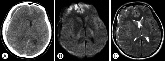
After returning to the general ward (about 20 min after the procedure), she complained of headache. Her mental status changed to drowsy in five min. Fifty min after the central catheter insertion, patient’s mentality became comatose with right pupil dilated to 6 mm. She was sent to CT room immediately with oxygen supply via ambu bagging. Her respiratory arrest occurred at CT room. Cardiopulmonary resuscitation was performed for 3 min which achieved spontaneous respiration. Brain CT revealed acute SDH on right frontotemporoparietal convexity (Fig. 1A).
The patient was immediately transferred to surgical intensive care unit and intubated. Mannitol and steroid were administered. When presented to neurosurgeon, neurological examination revealed fixed pupil of 6 mm/4 mm and Glasgow Coma Scale (GCS) score of E1VeM2. The patient showed decerebrated posture on pain. Brain magnetic resonance imaging (MRI) including diffusion weighted image was taken to rule out possible underlying parenchymal pathology and to estimate the damage from the herniation. In T2-weighted MRI, the hemorrhage appeared to be isointense mixed with hyper-intensity, indicating that the hematoma was a hyperacute stage (Fig. 1B). Fluid-attenuated inversion recovery image showed damaged frontal lobe from subfalcial herniation (Fig. 1C).
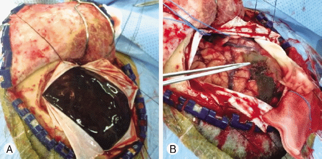
Emergency operation was performed about 4 hr after the patient felt the headache. After craniotomy, acute hematoma was suckable. Therefore, it was removed by suction (Fig. 2A). However, a piece of hematoma was stuck to the temporoparietal junction. After the removal of this last hematoma, blood pumped out from cortical artery. It assured us that the hematoma’s origin was this cortical artery (Fig. 2B). Right upon this observation, we alleged the radiologist that the hematoma is not related to C-line insertion. Craniotomy bone flap was removed and preserved at −70°C freezer for possible brain swelling. Removed hematoma underwent thorough pathological examination for possible hidden causative material. However, we found nothing but a hematoma.
After the operation, her mental status was improved to GCS of E1VeM4 at postoperative 3 weeks. Brain CT revealed a resolution of midline shift and decreased brain swelling.
The patient then underwent tracheostomy and cranioplasty. Three months after the craniotomy, her neurological status was partially recovered up to GCS of E2VtM4. She was then transferred to other hospital for rehabilitation.
DISCUSSION
Non-traumatic acute SDH are relatively frequent. There underlying causes include cerebral aneurysm, AVM, brain tumor, hematological disorders, and so on3,8). Spontaneous acute SDH of arterial origin is rare. It can be distinguished from other SDH by the following characteristics9). First, arterial bleeding is proved at operative finding or some other indirect evidence such as angiography. Second, the case must not be related to either traumatic history or known underlying vascular disease that could bleed itself such as arterial aneurysm, AVM, tumor, and coagulopathy. In addition, it should not be originated from other types of hemorrhage such as sub-arachnoid hemorrhage or intracerebral hemorrhage.
In this case, there was intraoperative finding of cortical arterial pumping. In CT, the hemorrhage showed as mixed formation with isodense and hyperdense lesion. In T2-weighted MRI, the hemorrhage appeared to be isointense mixed with hyperintensity, indicating that the hematoma was at hyper-acute stage. Considering the time of neurologic deterioration, it took only 30 min from starting headache to coma state, indicating that there was arterial bleeding.
Why arterial bleeding occurred in this case remains unclear. We have reviewed the central line insertion procedure. However, we did not find anything in the procedure at the left subclavian vein that could cause a cortical artery rupture of right cerebral hemisphere. Neither remarkable blood pressure change nor head trauma during the procedure occurred.
Several possible anatomical situations that might lead to cortical artery to rupture without traumatic events have been reported, including: (1) fragile arterial twig branching from cortical artery vulnerable to pressure connecting to dura mater, (2) a knuckle of cortical artery protruding through the arachnoid and adherent to the dura mater, (3) adhesions between a cortical artery and arachnoid or dura mater5). Intra-operative finding that suggesting one of them was not found in this case.
Spontaneous arterial bleeding related to hypertension2,10), alcohol abuse1,9) and the use of anticoagulant or antiplatelet agent have been reported. However, this patient did not have any underlying premedical history except thyroid cancer. In addition, her laboratory findings were within normal ranges.
CONCLUSION
Although arterial origin spontaneous SDH is rare, it has been reported in several case reports. Like other cases previously reported, the patient in this case report also had sudden neurologic deterioration and showed poor outcome. A cortical artery bleeding was found in operative field. Therefore, acute SDH without trauma history should be considered as an arterial bleeding. Early operation is recommended.