INTRODUCTION
Synovial cysts of the spine are cystic formations connected to the facet joint and contain synovial fluid17). These cysts are common in the lumbar spine, but are rare in the cervical spine17). Most cervical synovial cysts are found incidentally, but may cause radiculopathy or myelopathy due to compression of the spinal cord or nerve roots4). Generally, a symptomatic cervical synovial cyst is often associated with degeneration of the facet joints. Therefore, the pathologic mechanism involved in these cases is considered as of non-traumatic origin12). Few reported cases of a prior traumatic event being implicated in cyst enlargement due to acute bleeding into the cyst cavity, resulting in epidural compression.
We report a patient who presented with a hemorrhagic cervical synovial cyst without a definite trauma history that resulted in spinal cord compression. A review of hemorrhagic causes as a possible mechanism for cyst enlargement was also made.
CASE REPORT
A 74-year-old-woman with a 10-year history of lower back pain presented with a waxing and waning paresthesia and weakness of both legs that has slowly progressed over 1-year. The patient denied any recent traumatic event and the past clinical history was unremarkable, except for hypertension. The patient is not on any anticoagulation therapy. Initial neurological examination revealed G4/5 muscle power and diminished sensation in both legs. The patient denied any change in bowel or bladder function.
Magnetic resonance imaging (MRI) of the cervical spine revealed a heterogeneously enhanced cystic mass that has compressed the spinal cord posteriorly at the C7-T1 level. The extradural mass showed heterogeneous signal on T2-weighted images (WI) and the wall of the mass showed diffuse enhancement on contrast enhanced T1-WI. It was inseparable from the left C7 lamina and ligamentum flavum and was compressing the left posterolateral aspect of the spinal cord and the exiting left C8 nerve root (Fig. 1).
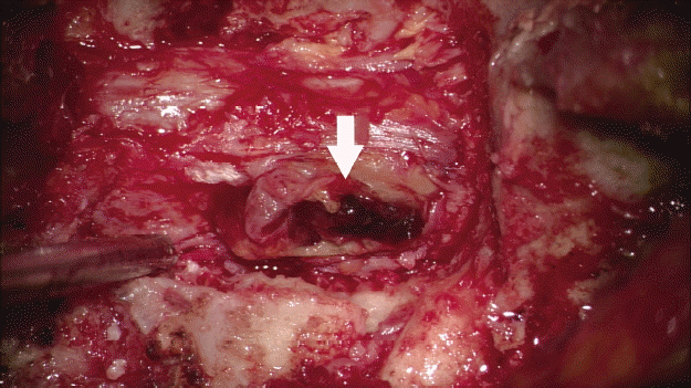
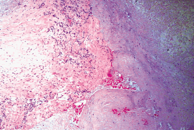
We performed a total laminectomy of C7-T1 and lateral mass screw insertion using a posterior approach with transpedicular screw insertion at the T1 level. The mass lesion was completely removed. The mass appeared in continuity with the left C7-T1 facet joint and was adherent to the dural sac. It had cystic components with intralesional subacute bleeding (Fig. 2). The histologic examination of the resected mass showed a cyst having thin fibrous wall with synovial lining, consistent with a synovial cyst (Fig. 3). The patient’s neurologic symptoms gradually improved over a few weeks after the surgery.
DISCUSSION
Spinal synovial cyst is defined as an extradural extrusion of the synovium through a capsular defect from a degenerate or unstable facet joint13). Based on histologic analysis, it is lined internally with a cuboid or pseudostratified columnar epithelium and contains clear fluid10). Since synovial cysts occur most frequently in the lumbar spine, symptomatic cases are much more commonly seen in those affecting the lumbar spine compared with the cervical spine14). The cause of synovial cysts in the cervical spine is multifactorial, but remains uncertain. It is thought that they start with facet joint degenerative change and erosion that extended through the wall of the joint capsule, triggered by hypermobility7,15) or due to a traumatic event15,16). Inflammatory factors may also play an important role in the development of synovial cysts, with upregulation of angiopoeitin-1, basic fibroblastic growth factor, substance P, platelet-derived growth factor, and interleukins at the site of the mechanically stressed facet joints contributing to synovial hyperplasia and leading to cyst formation15). Additionally, cervical synovial cysts have most commonly been reported at the cervicothoracic junction, suggesting a predilection for cyst development at a transitional junction between the mobile and fixed segments of the spine15).
The natural history of symptomatic cervical spine synovial cysts generally follow a slow, stepwise progression of symptoms and signs, all of which commence once there is compression of the spinal cord and/or of the exiting cervical nerve roots. The rapid deterioration of symptoms is thought to be due to hemorrhage into the synovial cysts, where trauma may play a role in the acute expansion9). The reason for hemorrhage into a facet cyst is unknown. Theories include increased venular vasculature of the degenerative synovium, as well as degenerative or traumatic rupture of the cyst itself18).
Computed tomography (CT) myelography and MRI are the most useful diagnostic investigations, delineating the synovial cyst arising extradurally next to degenerated facet joints1,3,5). Other useful radiologic findings are the presence of fluid within the cyst, and the extent of cyst compression of the spinal cord and/or of the exiting cervical nerve roots1,11).
Generally, surgery is indicated in the presence of severe pain and neurologic deficits. Some patients recover after surgical removal of the cyst, while others experience spontaneous resolution without surgery2,6). In lumbar synovial cysts, CT-guided percutaneous needle aspiration and/or corticosteroid injection have been attempted as non-surgical treatments, but these approaches have not been used to treat cervical synovial cyst8). In cervical synovial cysts, a percutaneous needle procedure may worsen the patient’s neurologic symptom because it may cause cyst rupture. Therefore, surgical excision is generally accepted as the treatment of choice for cervical synovial cysts because it results in early neurological improvement without the concern of recurrence12,15).
CONCLUSION
We present a case of lower cervical (C7-T1) synovial cyst with acute bleeding which lead to a sudden increase in the size of the lesion and spinal cord compression. The relative rarity of spinal synovial cysts and the even more limited number of cervical synovial cysts has led to very few reported cases. Surgery to evacuate the extradural hematoma and to excise the cyst can result in improvement or resolution of the neurological signs and symptoms.