INTRODUCTION
Dural arteriovenous fistula (dAVF) indicates the abnormal connection between the artery and vein in the dura and is known to comprise of 10% to 15% of arteriovenous malformations5). The most common sites of dAVF are the transverse sinus and the cavernous sinus, which are common in the West and East respectively3,5).
The treatment of dAVF includes compressing the internal carotid artery, radiation therapy, surgical therapy, and endovascular therapy. Currently, endovascular therapy is the most preferred treatment modality due to its high success rate and safety, in which transvenous embolization is commonly being performed with the path via the internal jugular vein (IJV) and inferior petrosal sinus (IPS) being used most advantageously6,15,16).
However, due to the failure of transvenous approach through femoral vein, the author would like to brief a case of dAVF embolization through superior ophthalmic vein (SOV).
CASE REPORT
A 72-year-old female admitted to our hospital presented with diplopia after falling down a month ago and exophthalmos which was aggravated 2 weeks ago. The patient was being treated for diabetes and Parkinson’s disease and did not have any other specific disease history. During the time of admission, the patient showed visual blurring and diplopia with pain in the left eye in which a bruit could be auscultated. The patient’s eye examination performed in the ophthalmology department showed restriction in lateral gaze, exophthalmos, and subconjunctival hemorrhage with visual acuity test showing 0.4 in the right eye and 0.3 in the left eye, and intraocular pressure of 17mmHg in the right eye and 25 in the left.
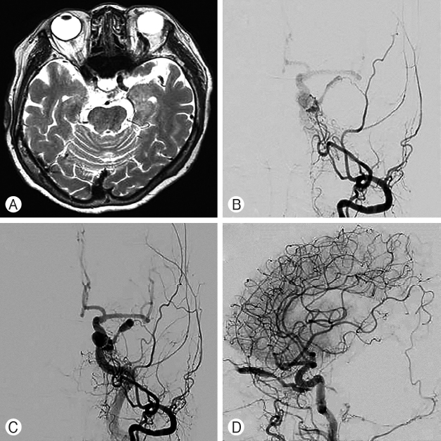
Brain magnetic resonance imaging performed after admitting to our department presented protrusion of the SOV and dAVF in the left cavernous sinus. Additionally, digital subtraction angiography (DSA) was performed using femoral artery in which a dAVF with blood supplied from the terminal of the maxillary artery and draining to the SOV and facial vein was observed (Fig. 1). Then, we planned the embolization 1 week later.
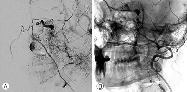
In order to treat the dAVF, embolization via the femoral vein, external jugular vein, and facial vein was attempted twice but the procedure failed as the microcatheter could not pass through the stenotic portion connecting the angular vein to the SOV (Fig. 2), therefore, due to the repeated failures, direct approach through the SOV was decided on. Embolization was performed after discussion on the exposure of the SOV with the ophthalmology and plastic surgery departments for the SOV approach.
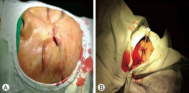
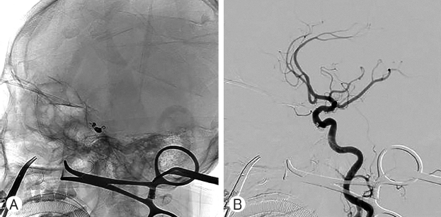
After general anesthesia, the SOV was exposed through an incision in the left eyelid. Temporary clipping with Yasargil mini curved clip was done, pediatric FAST-CathTM (4 FR, 8.5 cm; St. Jude Medical, Minnetonka, MN, USA) inserted (Fig. 3), and embolization performed by using pushable coils (TornadoR; Cook Inc., Bloomington, IN, USA; 3×2 8 EA) and detachable coils (AxiumTM Helix, Irvine, CA, USA; 2×6 2 EA, 2×4 1 EA) (Fig. 4). The patient was discharged without any particular neurologic symptoms and outpatient follow-up after 2 weeks showed normalized intraocular pressure and improvement of exophthalmos.
DISCUSSION
The dAVF is the abnormal vascular connection between the artery and vein arising from the dura, and is known to be common among the elderly, postmenopausal women with a history of hypertension, and secondary causes from trauma or surgery11). Characteristic symptoms include diplopia or ocular symptoms such as exophthalmos or ptosis which are common, and other symptoms such as headache or intracranial bruit7).
Treatment modalities for dAVF includes blocking the blood flow by clot formation in the cavernous sinus by compressing the ipsilateral internal jugular artery, surgical treatment, and endovascular treatment, in which the current method of preference is the endovascular treatment15,16).
The endovascular treatment method could be classified according to the method of approach into the transarterial approach and the transvenous approach. The transarterial approach is known to be difficult for complete embolization as there are many dural arteries and small branches, and has a high recurrence rate7). In comparison, the transvenous approach via the IJV and IPS through the femoral vein is most commonly used with the advantage of a high success rate in treatment compared to other modalities5,6).
If access to the IPS in the transvenous approach is unavailable, another approach through the pterygoid plexus, angular vein, and facial vein exists but there is a difficulty in treatment due to vessel curvature or hypoplasia, and stenosis8).
However, the approach through the SOV has a high success rate compared to other approaches and a low rate of complications, therefore being considered an alternative approach to the access via IPS through the femoral vein. The nonexistence of valves and the straightness of the vessel pathway are also advantages of the endovascular procedure using the SOV12).
Various case reports of endovascular embolizations through the SOV have been reported (Table 1). Reis et al.12) have performed endovascular embolization through the SOV on 85 patients based on previous study materials and stated that 76 among the 85 patients showed complete occlusion (of the malformation) which reached treatment success rate up to 95%. White et al.15) have reported on 8 patients in which the SOV approach was used for endovascular embolization as the transfemoral approach was not possible. Goldberg et al.4) have reported a case in which successful dAVF occlusion with improvement of eye sight and reduction of intraocular pressure were achieved, though the study also stated limitations of the unfeasibility of endovascular treatment procedure through the SOV if the vein is located deep within the orbit or not dilated.
Incision methods in the SOV approach consists of using the upper lid fold or incising under the eyebrow. There are studies suggesting that making an incision close to the superior orbit is favorable, and in our case, we determined the location of the SOV through angiography and performed skin incision by using the upper lid fold, which was the shortest route of access to the vein9,12). In the SOV approach, various complications such as hematoma or infection at the procedure site or the orbit, ophthalmic artery occlusion, and oculomotor palsy may occur, thus, a trained physician with an understanding of the accurate anatomical structures of the orbital area would be considered necessary7,10).
CONCLUSION
In cases of dAVF treatment where endovascular procedures through conventional routes are not feasible, endovascular procedure through the SOV should be considered, and in order to increase success rates and reduce complications, the exact location and exposure of the SOV through angiography should be performed as well as using a catheter with a narrow diameter and temporary clip which could be valuable for the procedure such as in our case.