Thoracic Outlet Syndrome
Article information
Abstract
Thoracic outlet syndrome (TOS) is a constellation of symptoms caused by the compression of neurovascular structures at the superior aperture of the thorax, properly referred to as the thoracic inlet. However, there is no consistent clinical presentation or definition. Based on a comprehensive literature review, this article presents the etiology, epidemiology, and clinical diagnostics of TOS, as well as the possibilities and outcomes of surgical treatment. The diagnosis and treatment are contentious, and some even question the existence of TOS. Its symptoms are often confused with those of distal compression neuropathies or cervical radiculopathies. The various surgical approaches for this syndrome are evaluated according to their ability to facilitate wide exposure, their potential morbidity, and their beneficial results. Most patients suffering from any form of TOS can benefit from surgical treatment. The duration of symptoms, socioeconomic factors and most notably, a stringent diagnostic workup and an adequate operative approach are important.
INTRODUCTION
Pain, weakness, and discomfort in the upper limbs are common. Diagnosis of these symptoms is often difficult because of the various pain-sensitive structures in the neck, the upper thoracic aperture, and the upper limbs. If other causes such as a cervical degenerative disease, rotator cuff rupture, tumors, peripheral nerve entrapment, and other neurologic diseases have been excluded, and the symptoms can be provoked during examination, the case is often classed under thoracic outlet syndrome (TOS). The term “thoracic outlet syndrome” was coined by Peet et al.19) in 1956 to describe several disorders attributed to mechanical compression of neural and/or vascular structures between the base of the neck and the axilla. TOS involves compression that results in injury or irritation of neurovascular structures as it passes through three narrow passages from base of the neck through the armpits to the arms. The most important of these is the interscalene triangle. The borders are the anterior scalene muscle anteriorly, the middle scalene muscle posteriorly and the medial surface of the first rib inferiorly. The triangle is small at rest but can be made smaller with certain provocative maneuvers. It can be further constricted by other structures such as fibrous bands, cervical ribs and abnormal muscles. The second passage is the costoclavicular triangle. The borders are the clavicle anteriorly, the first rib posteromedially and the upper border of the scapula posterolaterally. The third passage is the subcoracoid space beneath the coracoid process deep to the pectoralis minor tendon21).
ANATOMY
The thoracic outlet refers to the communication between the thoracic cavity and root of the neck. Three areas within the thoracic outlet where neurovascular compression can occur have been described: the interscalene triangle, the costoclavicular space, and the subpectoral tunnel. The most clinical important passage is the interscalene triangle, which is bordered by the anterior scalene muscle anteriorly, the middle scalene muscle posteriorly and the medial surface of the first rib inferiorly. This triangle includes the trunks of the brachial plexus and the subclavian artery. It should be noted that the subclavian vein flows anterior to the anterior scalene muscle. Immediately distal to the interscalene triangle, the neurovascular bundle enters the costoclavicular triangle, which is bordered anteriorly by the middle third of the clavicle, posteromedially by the first rib, and posterolaterally by the upper border of the scapula (Fig. 1). The neurovascular bundle then enters the subcoracoid space, also referred to as the retropectoralis minor space, beneath the coracoid process deep to the pectoralis minor tendon. Compression or irritation of the brachial plexus, or both, have been described within these spaces10).
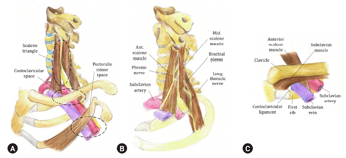
(A) An overall view of the left thoracic outlet, examined from the thoracic outlet area, showing that it is made up of several distinct spaces. Nerve (yellow) compression can occur at the scalene triangle or pectoralis minor space, whereas venous compression most often occurs at the costoclavicular space. Arterial injury is most often due to bone trauma at the scalene triangle. (B) Detailed view of the scalene triangle on the patient’s left side. (C) Anatomy of the subclavian vein at the costoclavicular space. [Modified from “Thoracic outlet syndrome: A common sequela of neckinjuries.”, by Sanders RJ, Haug CE, 1991, Lippincott, pp. 34, 236. Copyright 1991 by the Lippincott. Reprinted with permission].
PATHOANATOMY
1. First Rib Anomalies
These include bifurcation, synostosis with the second rib, and hypoplasia or absence of the first rib. First, rib hypoplasia is often associated with a postfixed plexus, which can be entrapped against a fibrous band representing the remnants of the rib (Fig. 2).

The cervical rib is a possible pathoanatomical factor of thoracic outlet syndrome (TOS). This anteroposterior cervical radiograph demonstrates a cervical rib that caused TOS in a 22-year-old female patient (white arrow).
2. Vertebral Abnormalities
The elongated transverse process of the seventh cervical vertebra was described by Raaf 20) to have an anomalous vertical fibrous band extending to the first rib. The band is located posterior to the brachial plexus, causing its compression. Other bony abnormalities associated with TOS include congenital pseudoarthrosis of the clavicle.
3. Cervical Muscles
The scalenus minimus muscle may cause neurovascular compression. The omohyoid muscle may also play a role in compressing the brachial plexus1). Telford and Mottershead29) described neurovascular compression caused by anomalous insertion of the scalenus anticus, medius muscles, or both. They restricted the term “scalenus anticus syndrome” to lesions in which this muscle is the sole etiologic factor. Based on anatomic dissections, they described two types of sharp tendinous insertions of the scalenus anticus that may contribute to neurovascular compression.
4. Fibrous or Muscular Bands
These soft tissue structures irritate or cause pressure on the brachial plexus but not the subclavian artery. Although very important in the cause of TOS, these fibromuscular bands are not detected with radiography but are often encountered at surgery24).
CLASSIFICATION AND SUBTYPES
1. Neurogenic TOS (NTOS)
Neurogenic types, accounting for more than 90% of TOS cases, were subdivided into “true” NTOS, and more common “disputed” NTOS. Both types are secondary to compression or traction injury to the lower trunk of the brachial plexus. True NTOS has named “true” because of the anatomical and electrodiagnostic evidence supporting the diagnosis. In contrast, the “disputed” NTOS, also known as nonspecific TOS, is believed by many to be an ambiguous clinical entity without objective clinical findings33). The classical form of true NTOS is called Gilliatt-Summer Hand8). This syndrome was first described in 1970 as thenar, hypothenar, and interossei weakness and/or atrophy, plus ulnar and medial antebrachial cutaneous hypoesthesia. Patients with true NTOS may report symptoms associated with pain, numbness, and weakness of the upper extremity. Overhead arm maneuvers or lift of heavy objects may aggravate the symptoms. However, approximately 85% of patients diagnosed with TOS have the disputed type. This form of TOS is associated with poorly defined and inconsistent symptomatology without objective evidence. As such, it has been embroiled in controversy, with many medical doctors questioning its very existence10). Patients with disputed NTOS will show similar symptoms of paresthesia and weakness with NTOS. But, usually there are more complaints of pain.
2. Vascular TOS (VTOS)
VTOS can be either arterial or venous. Venous compression can cause edema or cyanosis of the upper limb. It can also appear suddenly in the form of phlebitis occurring after varying degrees of effort. The patient can sometimes present only in the sequelae stage with thoracic collateral venous circulation. Arterial manifestations consist of upper extremity exertional ischemia or positional vasomotor dysfunction. Signs of vertebrobasilar insufficiency or Raynaud’s phenomenon can be observed due to compression of the origin of the vertebral artery22). The diagnosis of TOS is relatively simple when there are vascular symptoms in the upper limb when the arms are raised, but the vascular type is so rare that the venous type accounts for 2% to 3% of TOS and arterial type accounts for about 1%25). However, having vascular signs can help to guide the diagnosis in the presence of a predominantly neurological form of TOS.
DIAGNOSIS
1. Physical Examinations
1.1. Adson’s Test
The patient’s radial pulse is palpated (Fig. 3A), then the arm is externally rotated, extended and slightly abducted. The patient is asked to look towards the side being examined and to take a deep breath in (Fig. 3B). Abolition or a reduction in the radial pulse is a positive test.
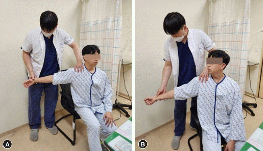
(A, B) Adson’s test.
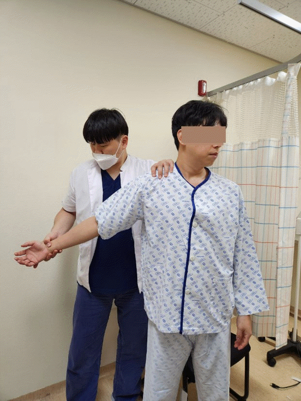
1.2. Wright’s Test (the hyper-abduction test)
The patient’s arm is abducted to 90 degrees in external rotation while palpating the radial pulse. Again abolition of the pulse suggests a positive test but there is a high false positive rate.
1.3. Roos’ Test
The patient’s shoulder is abducted and the elbow is flexed to 90 degrees. In this position the patient is asked to open and close their hands for three minutes (Fig. 4). Inability to complete this exercise pain-free or reproduction of presenting symptoms constitutes a positive result.
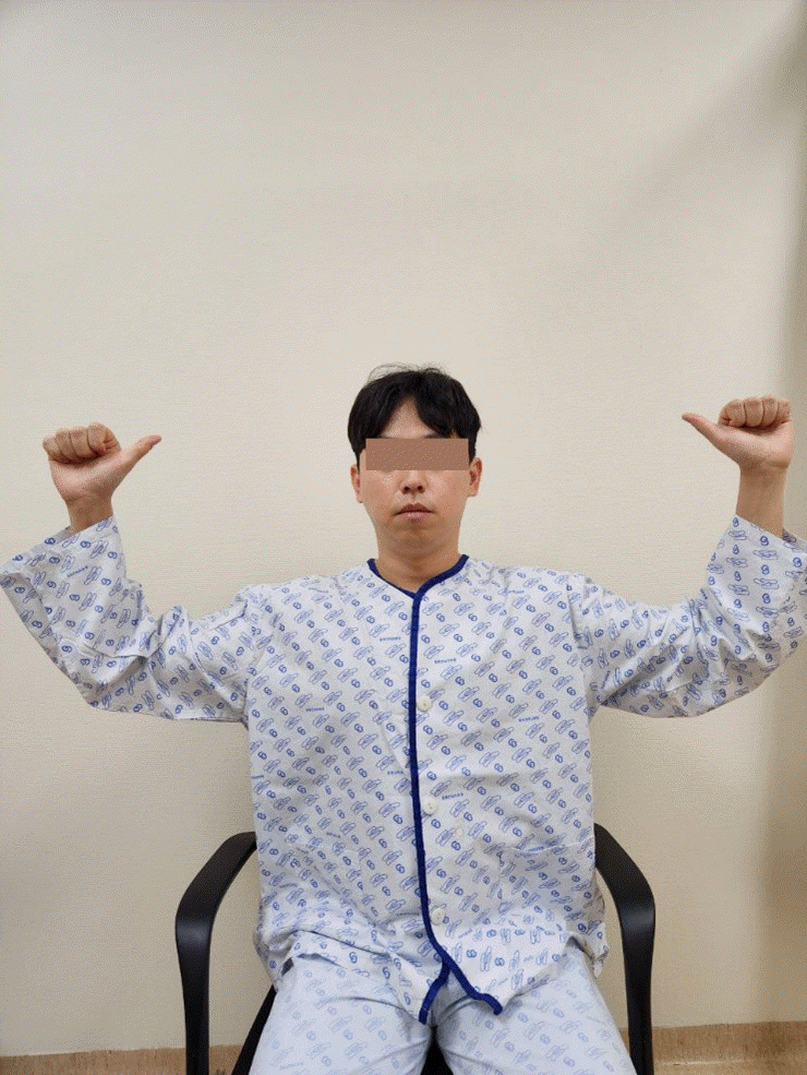
Roos’test.
2. Electro Diagnostic Studies
Diagnostic criteria for electro-diagnostic tests are clearly established in electrophysiological tests, particularly in true neurologic TOS, altered ulnar sensory conduction (low amplitude sensory nerve action potentials) and motor median conduction (low amplitude compound muscle action potential). Routine electro-diagnostic studies can sometimes yield normal results but when there is strong clinical suspicion of a NTOS and the classical electro-diagnostic abnormalities are not found, F waves may be used at rest and in provocative positions to help support the diagnosis11).
3. Other Diagnostic Tests
Scalene muscle block has been one of the most useful tests to confirm a diagnosis of NTOS. Although the response is subjective, a good response to the block has a high correlation with good responses to surgery for NTOS. Performed by the injection of a few mL of local anesthetic into the belly of the anterior scalene muscle, the test can be performed with electromyographic guidance or without it12,27). A neck X-ray is indicated to detect cervical ribs. Cervical magnetic resonance imaging or computed tomography scan is used to detect cervical spine disorders related to NTOS or may be a part of the differential diagnosis. Variations of magnetic resonance imaging are also being investigated for objective evidence of scalene muscle abnormalities or muscle compression of the brachial plexus. Magnetic resonance neurography is another recent imaging technique that shows deviations from the normal pathway by injecting a dye around the plexus6). Arteriography is not indicated in NTOS. As with the Adson test, demonstrating arterial compression is using an arterial sign to diagnose a neurologic condition. It is useless on NTOS. The only indication of arteriography is in ATOS, and then it is only necessary to plan the operation6). Venography is not indicated in NTOS. Venography is important in venous TOS and is indicated in patients with arm swelling and cyanosis. For VTOS, venography is more reliable than ultrasound3).
DIFFERENTIAL DIAGNOSIS
Clinical aspects of TOS are very wide, ranging from mild discomfort to severe symptoms14). Patients may also exhibit unilateral or bilateral signs or symptoms associated with a combination of nerve and vascular components9). Isolated vascular forms of TOS are more easily diagnosed but are also rare. So, the examiner must distinguish which symptoms are related to brachial plexus compression, which are vascular, and which are unrelated to the pathology of the thoracic outlet5). Cervical problems are more often characterized by persistent neck and shoulder pain, which may worsen depending on the position of the neck14). Distal compression neuropathy, such as carpal tunnel syndrome, is symptomatic alone and is predictable in relation to nerve distribution and exacerbated by wrist and elbow positions rather than shoulder or neck positions. The differential diagnoses important for the neurogenic form of TOS include musculoskeletal disease (such as arthritis or tendinitis) of the cervical column, shoulder girdle or arm, cervical radiculopathy or nerve compression of the upper extremities, idiopathic inflammation of the brachial plexus (also known as Parsonage-turner syndrome) and compression of the brachial plexus due to an infiltrative process or mass such as pulmonary apex Pancoast tumor7,16).
CONSERVATIVE MANAGEMENT
Preventive measures are essential to correct or eliminate identified hazards, particularly in the workplace, as discussed below18). The use of orthoses has also provided useful results for distal symptoms in some patients17). Rehabilitation has been performed for many years according to Peet’s protocol19), although a slightly modified protocol has often been used in recent years, consisting of an initial phase of analgesics and muscle relaxants, which appears to provide better results (particularly in painful neurological form). Correctly performed rehabilitation can provide long-term relief of symptoms in about two-thirds of patients. It is particularly effective for proximal pain28). In refractory forms, multidisciplinary management is essential along with evaluating the various factors participating in the maintenance of chronic pain and a retraining program such as those proposed for chronic low back pain15).
SURGICAL MANAGEMENT
Surgical intervention is considered in patients with vascular compression or NTOS that do not respond to conservative management. The three major surgical approaches to decompression of the thoracic outlet are transaxillary, supraclavicular, and posterior, although there are many variations and preferences that are surgeon dependent.
1. Transaxillary
First described by Roos23) in 1966, the transaxillary approach is the most commonly performed approach today. Proponents claim that it provides superior exposure for first rib resection, as well as for the removal of cervical ribs and fibrous bands, with a more cosmetic scar. In a review of TOS over 50 years, Urschel and Razzuk32), describe the transaxillary approach as an initial surgical approach through which they perform first rib and costoclavicular ligament resection, splenectomy, and C7, C8, and T1 neurolysis. They argue that while first rib resection can be accomplished via the supraclavicular approach, visualization is poor and requires retraction of the neurovascular structures.
2. Supraclavicular
The supraclavicular approach provides a more favorable exposure of the upper brachial plexus, the neck of the first rib, and the neurovascular structures. This approach is preferred by surgeons who perform isolated scalenectomies and removal of cervical ribs for NTOS13). Scalenectomy may be considered in patients with upper plexus-type NTOS, patients with TOS symptoms in the absence of abnormal bony architecture, patients who are excessively muscular or obese, or patients with recurrent TOS following prior first rib resection2). Although the supraclavicular approach may provide poorer exposure of the first rib compared with the transaxillary approach, Terzis and Kokkalis31) reported good outcomes and fewer complications with the supraclavicular approach for first rib resection. If arterial reconstruction is necessary, the supraclavicular approach is preferred30) (Fig. 6).
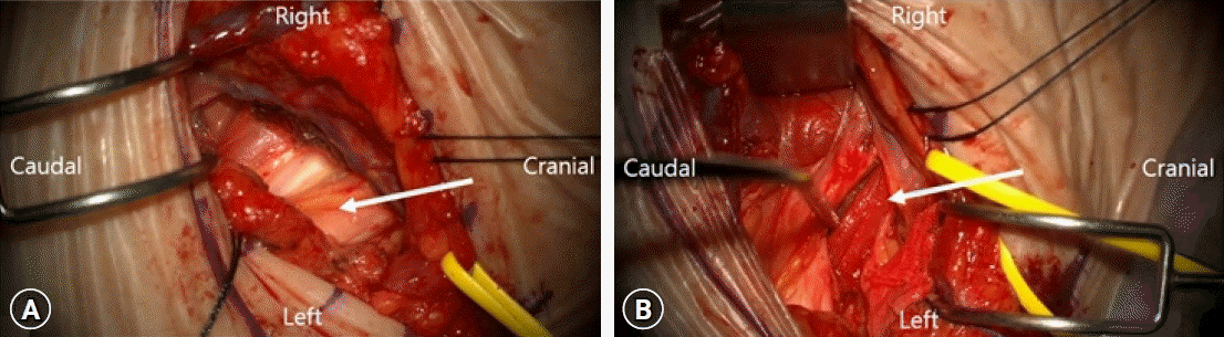
Left supraclavicular approach for anterior and middle scalene muscle resection for treatment of neurogenic thoracic outlet syndrome. (A) The exposed subclavian artery and brachial plexus (white arrow) after resection of anterior scalene muscle. (B) The middle scalene muscle (white arrow) behind the brachial plexus.
3. Posterior
The posterior approach, first described by Clagett4) in 1962, allows better exposure of the proximal component of the brachial plexus for neurolysis; however, the approach is more invasive and it can lead to postoperative shoulder morbidity and scapular winging26). Urschel and Razzuk32) reserve the posterior approach for removing rib remnants and performing brachial plexus neurolysis for patients with recurrent TOS.
CONCLUSION
A meticulous history and thorough physical examination are the most important factors for establishing a diagnosis of TOS. When indicated, radiographic and laboratory tests can improve diagnostic yield. Provocative positional maneuvers should be evaluated for vascular and neurological response. These maneuvers cannot make the diagnosis but can be a useful adjunct for confirming the diagnosis. VTOS is less common and often requires surgical treatment. NTOS is very common but less frequently requires surgical treatment. The selection of surgical approach and procedure must be determined by the nature of the pathologic condition and compression site.
Notes
No potential conflict of interest relevant to this article was reported.