De Novo Aneurysm of the Distal Middle Cerebral Artery and Subarachnoid Hemorrhage After Nasogastric Tube Insertion: A Case Report
Article information
Abstract
Distal middle cerebral artery aneurysm is rare and can develop from trauma, infection, atherosclerosis, or surgery. Nasogastric (NG) tube insertion is a common and safe procedure for stroke patients with dysphagia. In this case, the patient showed right internal border-zone infarction without major arterial occlusion. Magnetic resonance angiography of the brain showed no intracranial aneurysm. On the tenth day of hospitalization, we inserted an NG tube for feeding; insertion was difficult. Four hours later, the patients showed sudden mental deterioration. Computed tomography and computed tomographic angiography of the brain showed diffuse subarachnoid hemorrhage and ruptured de novo aneurysm at the right distal middle cerebral artery. Herein, we report a case of de novo aneurysm and subarachnoid hemorrhage that occurred after NG tube insertion.
INTRODUCTION
Distal middle cerebral artery (MCA) aneurysm is rare2,15). In a study by Dashti et al, among patients who underwent 1,012 surgical procedures for intracranial aneurysm, only four presented a distal MCA aneurysm2). The mechanism of distal MCA aneurysm formation remains unclear, but such aneurysm may occur after head trauma, infection, atherosclerosis, or intracranial surgery4,9,15).
Nasogastric (NG) tube insertion is commonly performed in acute stroke patients with dysphagia12). The procedure is performed by various medical staff, and is known to be generally safe1). However, NG tube insertion can be difficult and is associated with complications such as tube malpositioning, epistaxis, infection, local ulceration, perforation, feeding tube associated diarrhea, and aspiration pneumonia1,11). NG tube insertion also can increase intracranial pressure6). However, no case of intracranial hemorrhage has been reported.
Herein, we report a case of subarachnoid hemorrhage (SAH) that occurred after NG tube insertion, with the possibility of a newly developed aneurysm of the distal MCA. The patient did not yet have an aneurysm upon his initial admission, and had no risk factor of high blood pressure and intracranial pressure, except for the NG tube insertion. We try to discuss about the possibilities of suddenly increased intracranial pressure as the cause in the patient with ruptured de novo intracranial aneurysm after NG tube insertion.
CASE REPORT
A 76-year-old patient with a medical history of 20 years of hypertension and 10 years of diabetes mellitus was admitted in our institute with slurred speech and left facial palsy which occurred eight hours prior to admission. The patient’s consciousness level was alert. The patient’s Glasgow coma scale (GCS) was 15, and the patient’s National Institute of Health stroke scale (NIHSS) was 6. The patient’s motor grade was full in all extremities. All vital signs were normal: blood pressure, 110/70 mmHg; heart rate, 80 beats per minute; respiration rate, 20 breaths per minute; and body temperature, 36.5°C. The patient had dysphagia without a gag reflex. The patient had no trauma history and no family history of stroke or intracranial aneurysm. The patient’s initial brain CT scan showed a low-density lesion in the right frontal white matter, no intracranial hemorrhage, and no SAH. The patient’s brain MRI diffusion-weighted image series (DWI) presented a high signal intensity lesion in the right fronto-temporal lobe and the basal ganglia. These findings were concordant with the symptoms of acute cerebral infarction. The patient’s brain MRA showed no major arterial occlusion or intracranial aneurysm (Fig. 1, 2).
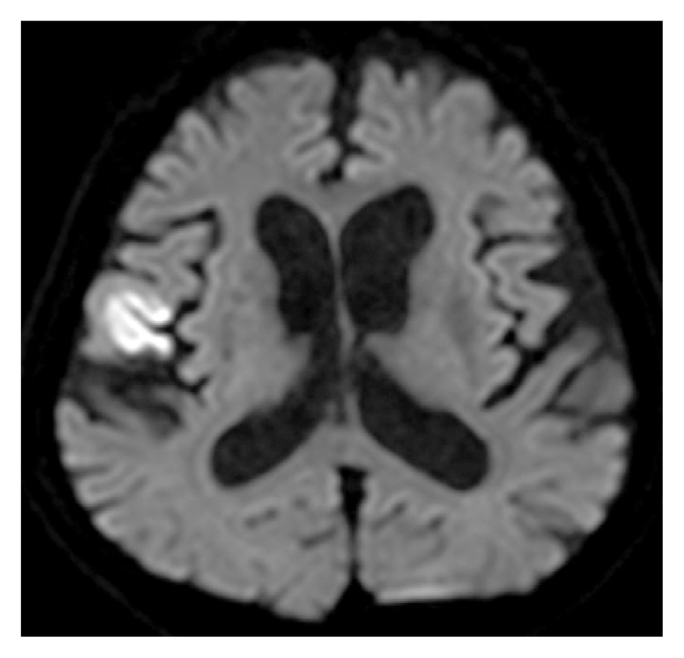
Initial brain MRI diffusion weighted image (DWI) showing a high-signal-intensity lesion in the right fronto-temporal lobe.
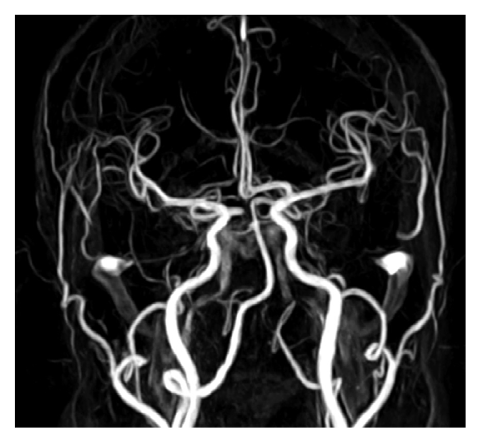
Initial brain MRA showing no intracranial aneurysm.
Intravenous tissue plasminogen activator (t-PA) was not applied, and intravenous ozagrel sodium 40 mg was started.
The patient had difficulty in swallowing, and an NG tube was inserted for his oral medication. During NG tube insertion, the patient was irritable and complained of severe pain. During the procedure, his blood pressure rose as high as 160/80 mmHg. After five hours, he vomited about 400 cc of vomitus with dark bloody content. The cause of his hematemesis was evaluated under sedation via endoscopy. Shallow esophageal ulcer due to irritation by the NG tube was observed in the esophagogastroduodenoscopy. We removed the NG tube and treated the patient with a proton pump inhibitor.
On the second day of the patient’s hospitalization, he had a fever above 38°C. His chest CT revealed aspiration pneumonia with right lower lobe haziness. The laboratory studies revealed white blood cell count of 14,000/uL (4,000–10,500/uL), erythrocyte sedimentation rate of 60mm/hr (0–20mm/hr) and C-reactive protein level of 15.80 mg/dL (0–0.3 mg/dL). The blood culture and sputum culture were negative. The patient was treated with the intravenous piperacilin sodium 4 g and tazobactam 0.5 g. His fever improved gradually over the next three days. The result of his follow-up examination was negative for pneumonia on the seventh day of his hospitalization.
On the 10th day of the patient’s hospitalization, the NG tube was inserted after consultation with a gastroenterologist for the anti-platelet medication. We tried to insert the NG tube repeatedly, but failed. After four hours, he suddenly became stuporous, and his GCS was 8. His brain CT showed thick subarachnoid hemorrhage and massive intracerebral hemorrhage in the right fronto-temporal lobe (Fig. 3). His brain CT angiogram revealed an 8×3 mm-sized aneurysm in the opercular segment of right MCA (Fig. 4). His family refused an emergency operation, and he expired after three days.
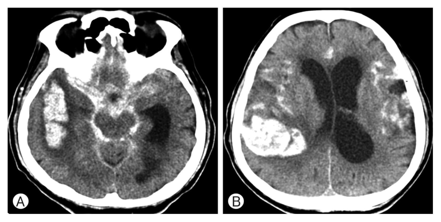
Brain CT at admission day 10 showing acute subarachnoid hemorrhage in both the sylvian fissures and the basal cistern (A), and intracerebral hemorrhage in the right frontotemporal lobe (B).

Brain CT angiogram at admission day 10 showing an 8×3 mm-sized, bean-shaped aneurysm at the opercular segment of right distal MCA (white arrow).
DISCUSSION
NG tube insertion ought to be performed carelessly, because fatal complications are uncommon. NG tube insertion can be distressing for patients, leading to increase in heart rate and systolic blood pressure5). NG tube insertion can also increase the intracranial pressure6). It may be associated with the start of an aneurysm17). Kurokawa et al. reported a case of a de novo aneurysm of the distal MCA which was caused by hemodynamic stress after superficial temporal artery-middle cerebral artery bypass surgery9). Hashimoto et al. reported that increased hemodynamic stress played a role in the growth and rupture of a distal posterior cerebral artery aneurysm after occlusion of the ipsilateral internal carotid artery7). In our patient, the enhanced brain CT revealed a cerebral aneurysm with high attenuation on the 10th day of his hospitalization. However, his initial enhanced T1-weighted brain MRI did not reveal any lesion suspected as an aneurysm at the same location (Fig. 5).

On the 10th day of the patient’s hospitalization, his enhanced brain CT revealed a cerebral aneurysm with high attenuation (A). However, his initial enhanced T1 weighted brain MRI did not reveal any lesion suspected as an aneurysm at the same location (B).
Mycotic aneurysms may be also associated with aneurysm formation. The distal MCA is the most common location of a mycotic aneurysm13). In this case, the patient had fever, aspiration pneumonia, and a distally located aneurysm, which may suggest a mycotic aneurysm8,10). However, the patient had fever only for three days, which subsided with antibiotic treatment. Blood and sputum cutures were all negative.
A sudden rise in blood pressure can cause intracranial aneurysm rupture16). It can be associated with intracranial hemorrhage14). In our patient, a sudden change in the blood pressure may have contributed to the rupture of the aneurysm in the distal MCA during NG tube insertion. Dolati et al. reported the formation and rupture of an anterior choroidal artery aneurysm associated with hemodynamic stress after ipsilateral posterior cerebral artery occlusion3).
There is an NG tube insertion technique that reduces the increase in the blood pressure. Dziewas et al. introduced the reflex placement technique that induced a swallowing reflex via water injection through another thin catheter during NG tube insertion5).
CONCLUSION
We experienced a rare case of a de novo aneurysm of the distal MCA which is thought to be associated with NG tube insertion. During the NG tube insertion, there was a sudden rise in the blood pressure, and SAH had occurred. However, it remains unclear whether the distal MCA aneurysm was formed as a result of infection or in association with the NG tube insertion.