Giant Intraosseous Cellular Schwannoma of the Sacro-pelvic Region in a Young Woman
Article information
Abstract
Giant intraosseous schwannoma is very rare. Schwannomas arising around the sacrum can reach a considerable size before becoming symptomatic because of their insidious and slow growth, which can allow the displacement of nerves and internal organs, often resulting in extensive bone erosion. The treatment of these giant schwannomas causes significant neurological disability. A 29-year-old woman was suffering from paresthesia of her left posterior thigh. Her symptom was aggravated since the last 5 months. Neurological examination showed paresthesia in the left S2 and S3 dermatome. There was no anal sphincter dysfunction or voiding difficulty. Plain radiography demonstrated foraminal widening of the left S2. Pelvic computed tomography (CT) and magnetic resonance images (MRI) showed a huge mass extending from the sacrum (S2) to the presacral area. The size of the lobulated mass was 4×4×6 cm. The patient underwent laminectomy and gross total removal of the tumor. Through a posterior approach, the tumor was removed without any neural or visceral organ injury, including the left S2 root. After a 71-month follow-up, the neurologic examination was normal, the urodynamic tests continued to show no abnormal signs, and the MRI studies revealed no evidence of residual tumor or recurrence.
INTRODUCTION
Cellular schwannoma is a benign variant of Schwann cell tumor, which is distinguished from classic schwannoma by vigorous cellular growth and absence of Verocay bodies. White et al.8) reported that the main sites of occurrence were the intraspinal and paraspinal region in the mediastinum and the retroperitoneum with the tumor sometimes occurring in the sacrum. Intraosseous schwannoma was reported to account for less than 0.2% of all bony tumors3), and sacral schwannoma accounts for less than 1% to 5% of all spinal schwannomas. Giant intraosseous schwannoma is very rare. The tumor often affects middle-aged women. As reported previously, schwannomas arising around the sacrum can reach a considerable size before becoming symptomatic because of their insidious and slow growth, which may allow the displacement of nerves and internal organs, often resulting in extensive bone erosion5). The treatment of these giant schwannomas causes significant neurological disability. In this paper, we showed that detailed preoperative planning, careful dissection and sparing of uninvolved nerve roots made it possible to prevent unnecessary neurological impairment.
CASE REPORT
A 29-year old woman was suffering from paresthesia of her left posterior thigh. Her symptom was aggravated since the last 5 months. Neurological examination showed paresthesia in the left S2 and S3 dermatome. There was no anal sphincter dysfunction or voiding difficulty.
Plain radiography demonstrated foraminal widening of the left S2. Pelvic computed tomography (CT) and magnetic resonance images (MRI) showed a huge mass extending from the sacrum (S2) to the pre-sacral area. The size of the lobulated mass was 4×4×6 cm and it was located above the anteflexed uterine corpus.
For planning of this surgical procedure, three-dimensional CT (3D CT) and MRI were beneficial in determining the extension and the nature of the tumor, but a digital subtraction angiography (DSA) study was not performed.
Through a linear shaped, sacral posteromedial incision, supplementary bone demolition, which is left hemilaminectomy of the sacrum, was performed. After a T-shaped dural incision was made, a whitish-yellow mass was noted under the dorsal root S2. The lesion was microsurgically debulked and removed. Via this posterior approach, we removed the tumor totally without any neural or visceral organ injury. Instrumental spondylodesis was not performed. Immediately after the procedure, the patient had no neurological deficit. On the postoperative radiographs, there was no sign of spinal instability. After a 71-month follow-up, the neurologic examination was normal, the urodynamic tests continued to show no abnormal signs, and the MRI revealed no evidence of residual tumor or recurrence.
DISCUSSION
Turk et al.7) reviewed 21 cases of giant intrasacral schwannoma among 79 cases of intraosseous schwannoma reported in the English literature until 1992. They stated that a giant intrasacral schwannoma was a rare retrorectal mass. Because of slow tumor progression, the patients experienced pain and swelling late during the development of the lesion. There have been no reported cases of malignant intrasacral schwannomas. However, it may be very likely for cellular schwannoma to deceive us into the wrong diagnosis of malignant peripheral nerve sheath tumor because of its remarkable growth and the invasion of bony tissue5). Malignant peripheral nerve sheath tumors of larger than 5 cm, especially those associated with neurofibromatosis, were reported to adversely affect the prognosis4). Therefore, biopsy should be performed preoperatively to establish a conclusive diagnosis8).
For the treatment of the tumor, total removal of the lesion was considered curative, and adjuvant chemotherapy or radiation therapy was not worthy of being carried out5,7,8). Turk et al.7) and White et al.8) emphasized that basically cellular schwannoma was benign and that unnecessary sacrifice of intact nerves should be avoided on removal of the tumor.
While planning sacral amputation, the proximal involved site in the sacrum may determine the level of sacral resection. For performing sacral amputation, it is important to know the resulting functional effects on the pelvic visceral organs and to know if the strength of the remaining pelvic girdle is enough for the normal load during activities of daily living10). Bilateral preservation of the S1 nerve roots is necessary to preserve the locomotor function of the lower extremities. Unilateral S2 and S3 nerve root preservation is expected to retain satisfactory urinary and rectal function. Regarding the biomechanical strength of the pelvic girdle, Gunterberg et al.2) described that an amputation done through the S1 vertebral body weakened the pelvic girdle by approximately 50% and an amputation between S1 and S2 weakened the posterior arch of the pelvis by approximately one third. Taking the above points into consideration, in our patient, high sacral amputation between S1 and S2 with marginal resection of the tumor and release of the left S2 and S3 nerve roots from the sacrum were performed. These surgical options were possible thanks to the development of a unique threadwire saw9). Furthermore, the lumbar spine was connected to the pelvis with spinal instrumentation accompanied by posterolateral fusion. Through a combined anterior and posterior approach, the large tumor could be removed en bloc. Our case underwent one-stage operation, which is different from the other case reports in which the operation was done by using a two-stage approach. During several months after surgery, the patient was bothered by difficulties with urination and defecation, but this impairment gradually improved within 7 months postoperatively. There were no signs of recurrence of the tumor. The clinical results indicated that the long time taken for the operation was worth it for achieving a satisfactory functional outcome, and cautious nerve root-sparing dissection may be the most important factor to lessen postoperative neurologic deficits.
Patients who have a large presacral schwannoma without any neurologic impairment, and no evidence for the tumor to change its size on imaging studies may not be candidates for a high sacral amputation because of the great possibility of sacrificing sphincter function or creating dreadful neurologic deficits intraoperatively. Another treatment option may be to simply observe the patients at the present moment. However, it must be kept in mind that the tumors have a potential to imperil bladder and bowel function and destroy the connection between the lumbar spine and pelvic arch if left untreated. Because the symptomatic recurrence after de-bulking or decompression by piecemeal dissection was reported to be very high in giant intrasacral schwannomas1,7), complete tumor resection will be able to yield enough content with the treatment to the patients eventually.
Preoperative imaging studies including radiographic films, CT (especially 3D CT), and MRI were beneficial in determining the extension and the nature of the tumor and are mandatory for planning the surgical treatment6). CT or MRI demonstrated well the association of the tumor to the neighboring visceral organs such as the rectum and the bladder. The preoperative angiographic study was valuable in identifying the arteries that fed the tumor. Although the tumor showed hypo-vascularity on the angiograms, fortunately embolization with coils done the day before surgery was useful to reduce blood loss because the arteries were undetectable and it was hard to secure hemostasis because the tumor occupied the entire cavity of the pelvis.
CONCLUSION
A case of intrasacral cellular schwannoma with one of the largest volumes previously described in the literature was reported. High sacral amputation after a combined anterior and posterior approach could be performed without causing any disability. Detailed preoperative planning, careful dissection and sparing of uninvolved nerve roots made it possible to prevent unnecessary neurologic impairment.
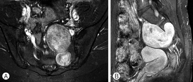
Preoperative contrast-enhanced axial (A), sagittal (B) Magnetic resonance images revealing a 4×4×6 cm sized huge schwannoma.

Preoperative plain radiograph demonstrated a widened foramen of S2 (A) Pelvic computed tomography also showed a presacral mass (B).

Postoperative enhanced magnetic resonance images showing that the gross total mass was removed (A) and photomicrographs obtained during histological analysis. Staining of the tumor (B) (H&E, original magnification ×60).