Pure Spinal Epidural Cavernous Hemangioma: A Case Report
Article information
Abstract
Pure spinal epidural cavernous hemangioma is difficult to differentiate from ruptured disc material and is rarely reported. It is usually reported in thoracic spine. The patient was a 73-year-old man suffering from chronic pain in the L5 dermatome area in his left leg. Computed tomography scans showed a slightly high-signal intensity epidural mass in lateral recess at the L4 vertebral body. This lesion showed hyper-intensity on T2-weighted magnetic resonance imaging (MRI) and enhancement on T1-weighted MRI with gadolinium enhancement. This mass-like lesion was surgically removed and the patient’s symptoms improved without complication. Here, we present a case of pure spinal epidural cavernous hemangioma, which should be included in the differential diagnosis of spinal epidural tumors. Early surgical treatment is important.
INTRODUCTION
Extradural cavernous hemangioma are very rare. In most cases, they arise from the vertebral bodies and extend into the extradural space6,9,15). Purely extradural cavernous hemangiomas without any vertebral body involvement are extremely rare, and account for only 4% of all extradural spinal tumors11,15). They are most commonly located in the posterior aspect of the thoracic epidural space and are rarely found in the lumbar space6,7,9,15). Clinically, they can cause symptoms of radiculopathy, which causes misdiagnosis as herniated intervertebral disc disease.
We present a rare case of a pure lumbar epidural cavernous hemangioma at the L4-5 level. This case is reported because of its rarity, the difficulty in making a preoperative diagnosis, and its favorable outcome after surgical treatment.
CASE REPORT
The patient was a 73-year-old man who had been suffering from left lateral thigh pain for the last 8 months; his symptoms were exacerbated within the past 3 months. He had progressive weakness in left lower extremity. Upon neurological examination, motor power in both lower extremities and deep tendon reflexes were intact. The patient had no past medical history and the laboratory tests were within normal limits except for mild hyperglycemia and the elevation in epinephrine (EPI; platelet function assay).
On the L-spine X-ray, endplate sclerotic change at L4-5-S1 were observed. L-spine computed tomography (CT) showed a ventral epidural mass around the lateral recess at the level of the L4 vertebra body. The mass was rounded in shape without calcification or acute hemorrhage (Fig. 1). From the L-spine magnetic resonance imaging (MRI), an epidural mass was seen as hyperintense on the T2-weighted images (WI) and isointense on T1-WI. Also, there was slightly heterogeneous staining on the gadolinium-enhancement T1-WI. Additionally, it was observed that the spinal cord was displaced to the left due to the mass (Fig. 2).

L-spine computed tomography showing a ventral epidural mass around the lateral recess at the L4 vertebra body level.

L-spine magnetic resonance imaging. An epidural mass was seen as isointense on T1-weighted images (T1-WI) and hyperintense on T2-WI. Also, there was slight heterogeneous staining on the gadolinium-enhancement T1-WI.
A L4 total laminectomy was performed and a dark red encapsulated mass with a soft consistency was found while the thecal sac was retracted. The mass was removed by en-bloc resection along with coagulation and no significant bleeding occurred during the operation. Neurological deterioration did not occur during the postoperative period. The patient’s left lateral thigh pain improved without any recurrence of symptoms until the current point, 3 months after the operation.
Histopathological examination revealed approximately 10 cc of pale pinkish fibrocartilaginous tissue and microscopic imaging allowed observation of the endothelial lining forming a cavernous channel, thus confirming cavernous hemangioma (Fig. 3).
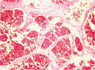
Microscopic image showing the endothelial lining, which formed a cavernous channel, thus confirming cavernous hemangioma.
DISCUSSION
Spinal epidural hemangiomas are rare lesions, constituting approximately 12% of cases of spinal cord vascular disease2,7). A pure epidural cavernous hemangioma was first reported in 1929 by Globus and Doshay. Cavernous hemangiomas are a collection of small capillaries covered with a single layer of endothelium, characterized by lobules, separated by fibrous connective tissue septa, and composed of irregular and dilated vascular channels4,14).
Cavernous hemangiomas are not real neoplasms. Clinical symptoms are caused by the mass effect, vascular movements, hemorrhage, thrombosis, cysts, or cavern formation. Cavernous hemangiomas may occur in any part of the body8). They mainly appear in the thoracic spine in an acute manner as the cord or conus are compressed2,3,5,12,14). Occurrence in the lumbar spine is very rare because nerve roots can tolerate compression better than the spinal cord; thus, it presents a chronic clinical course3,7). Pain does not resolve spontaneously, unlike in herniated intervertebral disc disease. For this reason, we need to consider cavernous hemangioma in patients suffering from chronic back pain or radicular pain which is not controlled by conservative treatment. If the diagnosis is a spinal cavernous hemangioma, only surgical resection can cure the problem.
As illustrated in our case, a purely epidural hemangioma, although uncommon, should be considered in the differential diagnosis of spinal epidural soft tissue masses. It is very difficult to diagnose before surgery. A study by Tekkök et al.13) reported on 14 surgical resections of cavernous hemangioma, none of which was diagnosed preoperatively.
MRI is the most useful method of diagnosing a cavernous hemangioma. It usually shows an isointense T1-WI and a hyperintense T2-WI. It often shows irregular or lobulated enhancement, and this needs to be differentiated from other diseases like intervertebral disc herniation, schwannoma, neurofibroma, angiolipoma, osteochondroma, synovial cyst, lymphoma, chordoma, and Ewing’s sarcoma. For example, intervertebral disc herniation may present with none or peripheral enhancement and schwannoma may present less enhanced or neural foraminal widening. Whereas lymphoma presents as isointense on T2-WI. In the case of angiolipoma, hyperintensity on T1-WI can be observed owing to its high fat content1,4–14).
Cavernous hemangiomas have a tendency to bleed and grow, so early surgical excision is recommended as soon as possible14). However, they are hypervascular lesions, so massive bleeding can be seen during the operation. En-bloc resection after coagulation is preferred4,6,14). As in our case, most patients show a good prognosis and an improvement in their symptoms after surgical resection of a cavernous hemangioma1,4,7,10,12).
CONCLUSION
Pure spinal epidural cavernous hemangioma is very rare. It can cause radiculopathy like intervertebral disc herniation, which makes the condition difficult to diagnosis preoperatively. MRI is a useful tool in helping to differentiate it from other diseases, and early surgical treatment is important.