INTRODUCTION
Pyogenic spondylitis accounts for 2% to 4% of all musculoskeletal infections14). Despite advancements in antibiotics and medical techniques, the incidence of spinal infections is on the rise, likely secondary to a growing elderly population with chronic diseases and a rise in drug abuse17). Pyogenic spondylitis is primarily caused by Gram-positive aerobic microorganisms—specifically Staphylococcus aureus (S. aureus)—with a reported incidence of up to 80%13). Anaerobic spondylitis, however, is exceedingly rare, constituting <3% of all pyogenic spondylitis cases10). Parvimonas micra (P. micra), an anaerobic Gram-positive coccus formerly known as Peptostreptococcus micros and Micromonas micros, has been identified in only a handful of cases worldwide—one reported in Korea in 20127,15). This case report describes an extremely rare paraspinal abscess and pyogenic spondylitis caused by P. micra following dental extraction in a patient without any specific risk factors. This study was approved by the Institutional Review Board of Pusan National University Yangsan Hospital (approval no. 55-2023-059).
CASE REPORT
A 71-year-old male patient presented to our hospital's emergency room with a three-day history of new-onset acute low back pain and radiating pain from the right hip to the right leg. He reportedly underwent acupuncture treatment at a traditional medicine clinic one month prior for back pain and periodic right leg paresthesias. He experienced fever-like symptoms three days before hospital admission but was afebrile upon examination. Physical examination showed marked tenderness in the lower lumbar region and pain-induced limited mobility, with no leg weakness. His past medical history was significant for hypertension, diabetes mellitus, and dyslipidemia. He did not use steroids or immunosuppressive drugs.
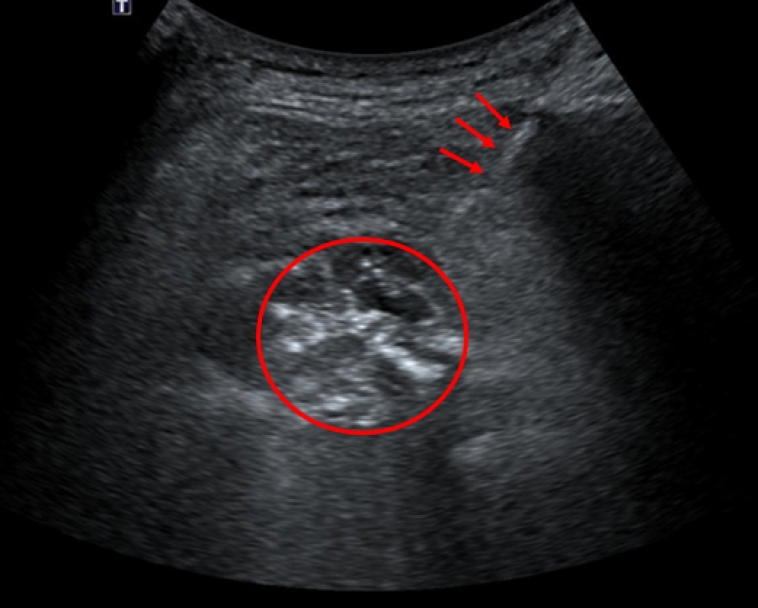
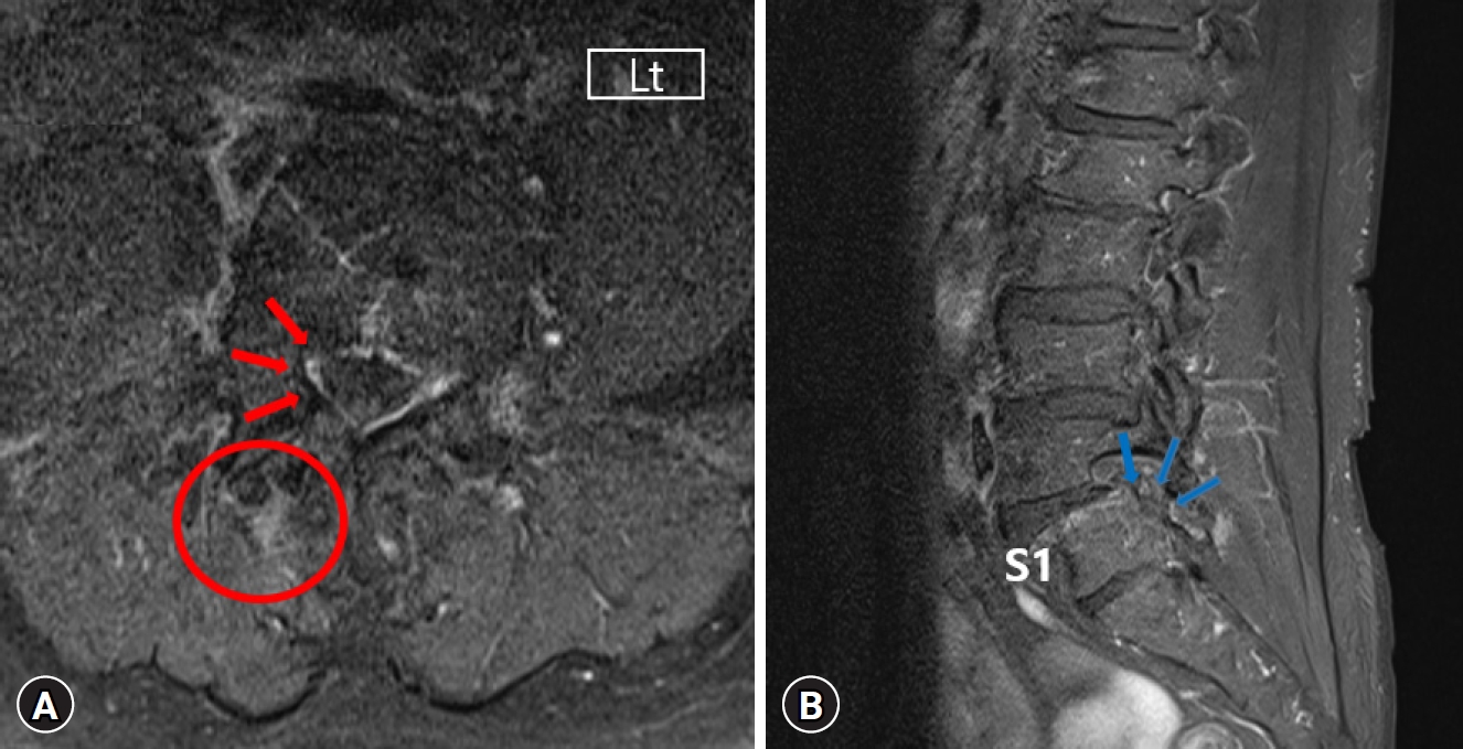
Initial laboratory findings revealed an elevated C-reactive protein (CRP) level of 2.60 mg/dL and a white blood cell count of 10.36 cells/mm3 with a neutrophil predominance of 69.3%. Liver and kidney function tests, urinalysis, and serologic tests for hepatitis B, C, and human immunodeficiency virus were normal. Considering his recent acupuncture treatment, we suspected a spinal infection. Contrast-enhanced magnetic resonance imaging showed suspected pyogenic spondylitis in S1 body and an epidural abscess at L5/S1 level, with a smaller abscess (1.0 × 1.7 cm) in the right paraspinal muscle (Fig. 1). Because we did not suspect epidural compression necessitating surgery, we initially pursued a conservative approach. Blood cultures and an ultrasound-guided biopsy (Fig. 2) were conducted for bacterial identification before antibiotic therapy. Empirical treatment with cefazolin (2 g every 8 hr) commenced, targeting skin flora.
When the culture results identified Gram-positive diplococci in both the blood and abscess (Fig. 3), we switched the patient to ampicillin/sulbactam after consultation with the Infectious disease specialist. P. micra, typically associated with dental plaque and chronic periodontitis, was confirmed to be the causative agent. The patient's recent dental history included tooth extraction for implant placement one week before hospital admission. Post-extraction, he reportedly experienced oral pain, gingival swelling, and fever, suggesting an oral origin of the spinal infection.
The patient responded well to the antibiotic regimen, and his CRP level was significantly reduced to 0.69 by hospital day 7. This downward trend continued as his symptoms—including back and radiating pain in his right lower leg pain—also improved. By hospital day 13, his CRP level had normalized to 0.19 and remained within the normal range. Given the patient's favorable response to conservative management, we deemed surgery unnecessary at this time.
Despite the common practice of switching from intravenous to oral antibiotics after 4 weeks in patients with pyogenic spondylitis, this patient opted to continue with a full 6-week course of intravenous antibiotics. Afterward, he was discharged on oral amoxicillin at a dose of 1 g three times daily. He continued to improve during outpatient follow-up, with no pain recurrence (Fig. 4). His laboratory test results also normalized, indicating successful infection management and resolution of the pyogenic spondylitis and associated abscess, as illustrated in Fig. 5.
DISCUSSION
Pyogenic spondylitis is an inflammatory condition that affects the spine and is characterized by abscess formation, edema, and destruction of bone tissue. S. aureus is the most common causative pathogen4). Pyogenic spondylitis from anaerobic bacteria, although accounting for less than 3% of cases, can also produce severe complications13). The pathogenic organisms frequently isolated in anaerobic spondylitis are Bacteroides, Propionibacterium acnes, and Peptococcus. Such infections caused by anaerobic bacteria often arise post-surgery, from direct trauma, from the spreading of adjacent soft tissue infections, or hematogenous distant infection sites12). Those with weakened immune systems—such as individuals with chronic diseases, intravenous drug users, or patients undergoing immunosuppressive therapy—are particularly vulnerable to anaerobic spondylitis2).
P. micra is an anaerobic Gram-positive coccus, previously named Peptostreptococcus micros or Micromonas micros, that exists as a normal flora on human skin, around teeth, or in the vagina; however, it is most commonly found in the oral cavity. When pathogenic, especially in the oral cavity, P. micra is implicated in periodontal diseases like gingivitis and periodontitis and systemic infections such as endocarditis, especially in immunocompromised individuals. A patient with pyogenic spondylitis caused by P. micra, complicated by infective endocarditis, has been reported in immunocompromised individuals. However, in the present case, there was no clinical evidence or history of immunosuppressant use, so it was decided not to perform an initial echocardiogram following consultation with an infectious disease specialist during the diagnostic phase3). When compared to infections caused by other organisms, pyogenic spondylitis due to P. micra has demonstrated a more favorable clinical outcome. The exact cause for this observation is not yet clear; however, it is hypothesized that the higher susceptibility to antibiotics compared to other organisms may contribute to this outcome. Additionally, clinical symptoms associated with P. micra-related pyogenic spondylitis are often indistinct, with a slow progression. Identification of P. micra can be challenging as it may require specific culture media or methods. Consequently, P. micra infections were historically underdiagnosed, but recent advancements in diagnostic methods have led to more frequent reporting of infections caused by P. micra1).
Our patient’s history of acupuncture was initially considered a potential causative factor. However, cultures eventually identified P. micra as the pathogen, and further past medical history-taking revealed that the patient had a tooth extraction for implant placement one week before admission. The ensuing symptoms of fever and extraction site swelling and pain aligned with the diagnosis and indicated that the dental procedure likely precipitated bacteremia, leading to pyogenic spondylitis.
The treatment protocol for pyogenic spondylitis, particularly about antibiotic selection and duration, is largely empirical due to limited research13). Standard practice involves administering high-dose intravenous antibiotics for 4 to 6 weeks, then transitioning to oral antibiotics until full recovery5). It is common to switch from intravenous to oral therapy at around four weeks; however, shorter periods of intravenous treatment are associated with an increased risk of therapeutic failure9). P. micra, like other strictly anaerobic organisms, proliferates in low-oxygen environments, making it challenging to culture in a laboratory setting due to its specific growth requirements. Consequently, specific techniques for confirming anaerobic organisms are necessary for identifying P. micra from clinical samples. Available diagnostic tests for P. micra include microarray, 16S rRNA sequencing, and matrix assisted laser desorption/ionization time-of-flight mass spectrometry (MALDI-TOF MS)16).
Susceptibility testing for anaerobic bacteria is complex and is not routinely performed. In the case of our patient, susceptibility testing was not conducted. Antibiotic treatment for spinal abscesses and spondylitis caused by anaerobic bacteria is not established due to the rarity of such cases8). According to previous studies, we administered ampicillin/sulbactam for P. micra7). Beta-lactam antibiotics, such as penicillin and carbapenems, are effective against anaerobic Gram-positive cocci6). Penicillin is generally considered the most appropriate treatment for Peptostreptococcal infections; however, resistance to P. micra has been reported15). The ideal treatment duration is not established but reportedly ranges from 2 to 10 weeks11). Our patient's significant improvement after one week of antibiotic therapy indicated a positive clinical outcome, as evidenced by reduced pain and normalization of CRP levels.
Indeed, anaerobic pyogenic spondylitis frequently presents clinically as persistent back pain, and fever may be absent in about half of the cases, complicating diagnosis. Blood cultures are crucial for identifying the causative organism in suspected bloodstream-disseminated anaerobic infections. Direct lesion sampling for culturing can guide targeted antibiotic therapy. While S. aureus is prevalent in pyogenic spondylitis, a broad differential diagnosis that includes anaerobes is vital. A thorough patient history is essential, revealing potential infection sources like recent surgeries, invasive procedures, or dental work. In this case, detailed history-taking linked the infection to a dental extraction and subsequent P. micra bacteremia, emphasizing the importance of comprehensive diagnostic processes.
CONCLUSION
The case underscores the rare yet potentially serious, diagnosis of pyogenic spondylitis caused by anaerobic bacteria, particularly P. micra. The severe nature of such infections emphasizes the importance of culture testing for precise diagnosis and effective treatment. This case further highlights the crucial role of comprehensive patient history in revealing vital details like recent dental procedures. In the case of our patient, this information provided essential insights into the infection’s source. Such attention to detail in clinical evaluation ensures that less obvious— but clinically significant—causative factors are not missed.