INTRODUCTION
The pathophysiology of traumatic SDG remains somewhat unclear. This case shows a rare etiology of traumatic SDG and demonstrates the usefulness of CT cisternography to determine cerebrospinal fluid leakage from an implant.
CASE REPORT
A 68-year-old male patient visited the emergency department after a passerby found him lying on the road with a bicycle. His initial mental status was a stupor mentality (Glasgow Coma Scale [GCS] score, GCS score 10). He was diagnosed with neurolymphomatosis in 2006. For chemotherapy, he had an Ommaya reservoir targeted to the right lateral ventricle via the right Kocher’s point.
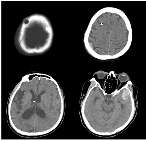
To identify the cause of his altered mentality, a brain-computed tomography (CT) was performed. Multiple traumatic injuries were identified. There was a hemorrhagic contusion at the left temporal lobe, an epidural hematoma at the right parietal area, a left tentorial subdural hematoma, and a right frontoparietal skull fracture (Fig. 1).
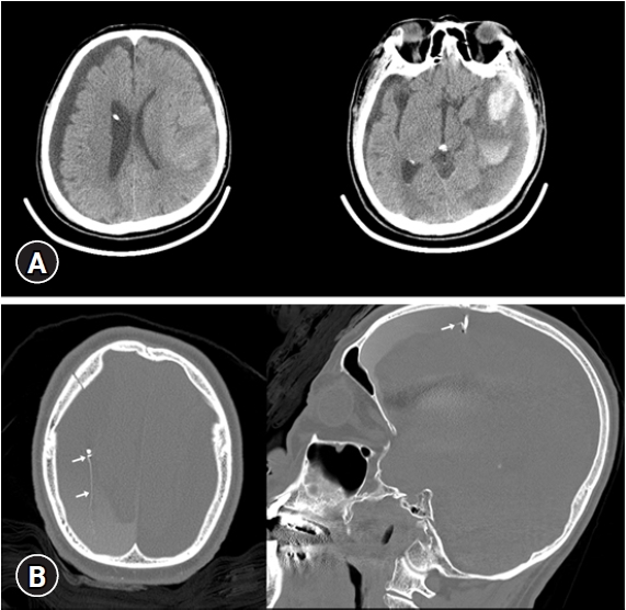
Seven days after admission to the intensive care unit, a bilateral subdural hygroma (SDG) was identified on brain CT (Fig. 2A). The amount of hygroma was dominant on the right side. Considering cerebrospinal fluid (CSF) leakage at the spine level due to trauma, a magnetic resonance (MR) myelography was performed, but there was no evidence of CSF leakage.
We focused on the Ommaya reservoir as the etiology of rapid SDG formation because of the adjacent skull fracture. We assumed that CSF leakage could develop from the damaged Ommaya reservoir by trauma. Therefore, we performed CT cisternography. The contrast was directly injected into the Ommaya reservoir and CT images were obtained. In the cisternography, the contrast leakage points were identified from the Ommaya reservoir (Fig. 2B).
We decided to remove the Ommaya reservoir and perform external drainage of the SDG by burr hole trephination simultaneously. After the procedure, the SDG seemed to be decreased slightly. However, a follow-up CT image showed that there was no significant improvement.
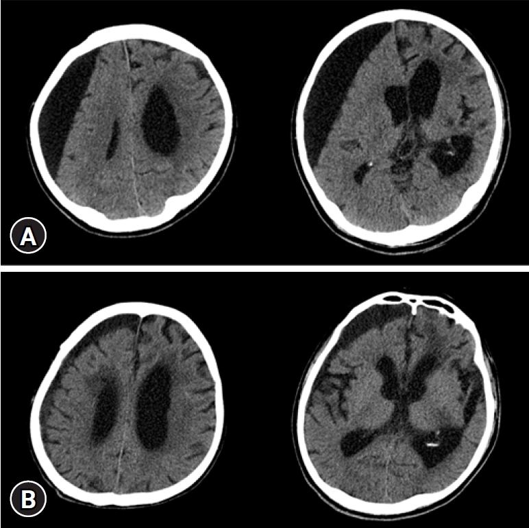
Finally, the case underwent surgical exploration with craniotomy. In the surgical field, there was a permanent tract from the ventricle to the subdural space, which was the remnant of the previous Ommaya reservoir. We occluded it with Absorbable Gelatin Sponge (Gelfoam; Pfizer, New York, USA) and fibrin sealant patch (TachoSil; Baxter, Deerfield, IL, USA). One month after surgery, the SDG decreased significantly (Fig. 3).
DISCUSSION
A SDG is defined as an acute or chronic accumulation in the subdural space of the CSF, often of altered composition. A major cause of SDGs is trauma2).
The pathophysiology of traumatic SDG remains somewhat unclear. The current theory relates to the tearing or destruction of the arachnoid membrane resulting in a one-way flap, which causes CSF leakage that accumulates into the subdural space and prevents reabsorption3,6). Therefore, we generally cannot find the exact focus of SDG macroscopically. The treatment of SDG is also controversial due to the uncertain pathophysiology and difficulty identifying the leakage point1).
In this case, we identified the exact origin of SDG formation. The first was the damage to the Ommaya reservoir and the second was the permanent tract from the ventricle to the subdural space due to the Ommaya reservoir. Furthermore, we demonstrated the leakage focus of CSF visually with CT cisternography.
CT cisternography is usually used to find CSF otorrhea or rhinorrhea4). Its CT cisternography is usually used to find CSF otorrhea or rhinorrhea4). Images are obtained by injecting contrast after a lumbar puncture, but it can be implemented through another accessible route to the subarachnoid space or ventricle. We injected the contrast directly into the Ommaya reservoir and obtained a CT image to determine whether the CSF leakage was present in the Ommaya reservoir. Also, compared to MR myelography or radionuclide cisternography, this procedure obtains more precise images5). Therefore, if it is difficult to identify a clear CSF leakage point using a different modality. CT cisternography using high-resolution CT is of great help.